How can sound waves heat a small target deep inside the body while passing through tissue on the way there? Focused ultrasound uses waves that converge at a chosen region, concentrating acoustic energy there. The idea resembles using a lens to bring light to a bright point, though sound travels through tissue by a different mechanism and its path must be carefully controlled.
Sound carries mechanical energy
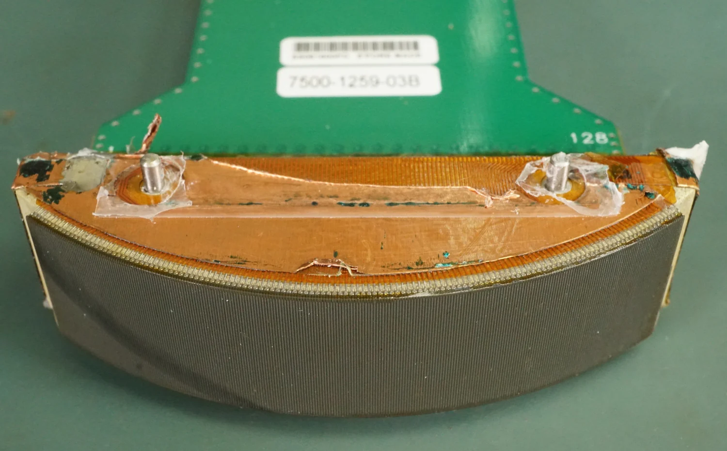
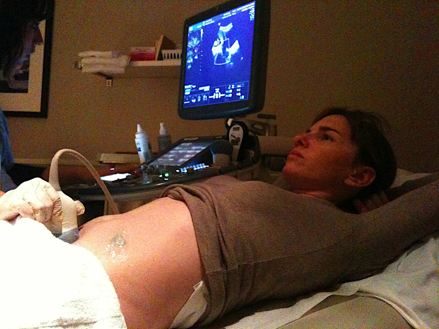
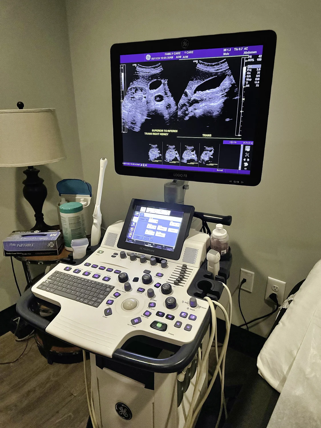
Ultrasound is sound at frequencies above the range of human hearing. A transducer converts electrical energy into sound waves and can also detect returning echoes. In diagnostic imaging, echoes from boundaries between tissues are timed to estimate their distance and build an image [1]. Therapeutic ultrasound uses sound energy to interact with tissue rather than primarily to form a picture.
A focused beam is created so that waves arriving from different parts of a transducer reinforce one another in a small region. Away from that focus, the energy is distributed across a wider path; at the focus, it becomes concentrated. The result depends on the beam, tissue properties, and the ability to locate the target. In high-intensity focused ultrasound (HIFU), the goal can be to heat or destroy selected tissue [1].
A focal point concentrates effects
Picture ripples on a pond meeting at one spot. Where wave crests reinforce each other, the resulting disturbance is larger. In tissue, ultrasound waves are pressure variations that cause microscopic motion. At sufficiently high intensity, this mechanical energy can become heat. Heating is not the only possible effect of therapeutic ultrasound, but it is a central mechanism in many focused applications.
Hyperthermia is a treatment approach that uses heat to damage cancer cells, usually alongside other treatments such as radiation or chemotherapy [2]. Ultrasound can be one way of producing heat in tissue. The temperature reached and the volume affected matter: too broad a heated region could harm healthy tissue, while a misplaced focus might miss the intended target. These are reasons therapeutic systems use imaging and monitoring to guide treatment.
Imaging and treatment have different jobs
Diagnostic ultrasound uses relatively low-intensity waves to gather echoes and produce images. Therapeutic ultrasound uses higher acoustic output to modify or destroy tissue [1]. The same broad technology therefore serves two different purposes: one measures, the other acts. A scan image is not evidence that tissue has been therapeutically changed, just as a treatment beam is not necessarily producing a diagnostic picture.
Ultrasound or MRI may be used to locate a target and guide HIFU in real time, as well as to assess treatment effects [1]. MRI uses magnetic fields and radio waves rather than ionizing X-rays; it does not use plasma. These distinctions matter because the guidance method and the energy-delivery method are separate parts of the procedure.
Applications require evidence of their own
Focused ultrasound is used or studied for different purposes, and the evidence and availability vary by application. NIBIB describes HIFU as being investigated for modifying or destroying abnormal tissue and lists specific approved uses, including uterine fibroids, pain from bone metastases, and prostate tissue ablation [1]. The existence of one established use does not establish benefit for every proposed target or condition.
Other research directions include breaking up clots, delivering drugs to selected locations, and temporarily opening the blood-brain barrier [1]. These are distinct goals with distinct physical and biological challenges. The basic physics explains how acoustic energy can be concentrated; it does not by itself show that a particular treatment improves outcomes. Separating mechanism from clinical evidence lets us appreciate the engineering while keeping claims about benefit tied to the specific application.
Explore the physics: an explanatory diagram

Sources & further reading
- Ultrasound NIBIB
- Hyperthermia to Treat Cancer National Cancer Institute
Prepared for the independent PSI Center reading library. About edition dates and source checking.
This explainer describes scientific principles. It does not assess individual treatment choices.