What happens to a sound beam when it meets skin, fluid, muscle, or bone? For a medical device designer, that question can be explored first in a model: a mathematical description of how sound moves and reflects through materials. Acoustic models help compare designs and predict where energy may go, while physical experiments and clinical evidence remain necessary to judge performance.
Sound becomes a measurable signal
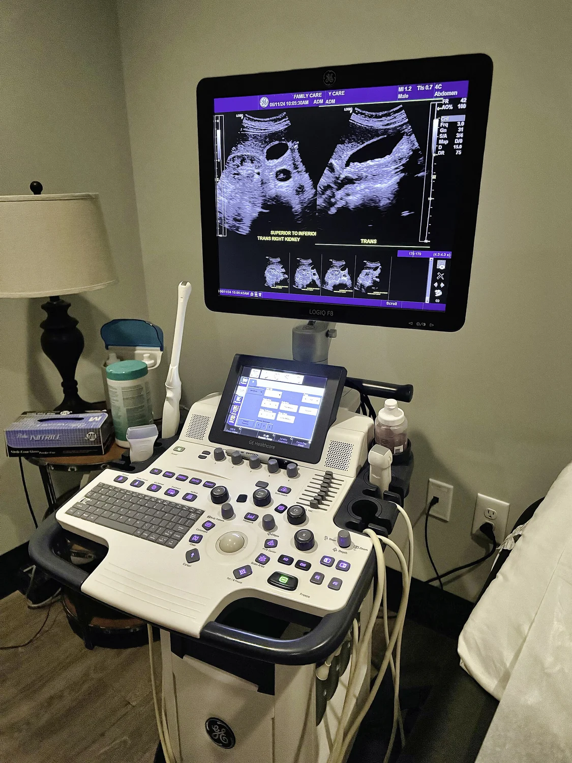
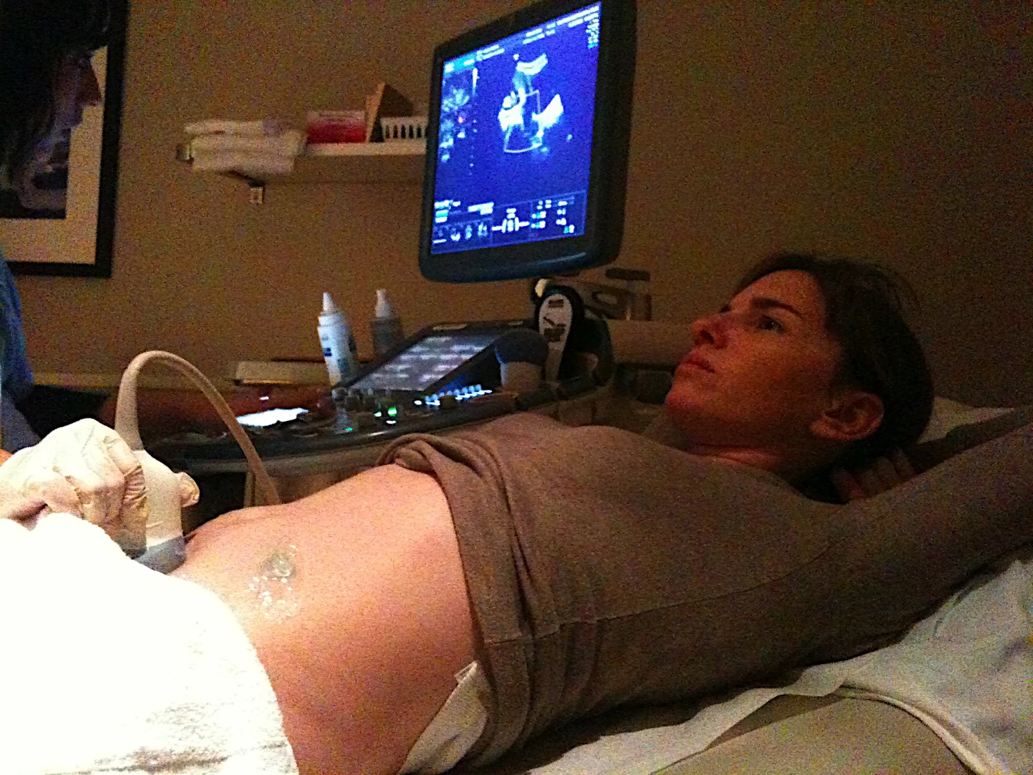
Medical ultrasound uses sound waves above the range of human hearing. A transducer—an instrument that both emits and detects sound—sends pulses into the body. At boundaries between tissues, some sound reflects back. The returning echoes become electrical signals, and the scanner estimates the distance to a boundary from the time each echo takes to return [2].
A simple mental experiment is to shout across a room and listen for an echo. The delay hints at how far away a wall is. In tissue, the device uses many short pulses and reflected signals to assemble a two-dimensional image. The same basic physics—sound traveling, interacting with boundaries, and returning—can be represented in equations for design and simulation.
A model tests design choices
Computational modeling uses computers to simulate complex systems through mathematics, physics, and computer science. A model can represent variables such as material properties, geometry, and operating conditions, then calculate predicted outputs under different scenarios [1]. For an acoustic device, designers might compare how changing a probe shape or target position affects a modeled sound field.
This is useful because a computer can run many virtual experiments before researchers select a smaller number for laboratory testing. Mechanistic models encode known physical principles; data-driven models identify patterns in measured data. Hybrid approaches combine both, but every prediction is limited by the assumptions, input data, and range of conditions the model represents [1].
From image formation to tissue interaction
For diagnostic ultrasound, the goal is to turn echoes into information about anatomy or function. For example, Doppler ultrasound uses changes in returned sound to visualize blood flow, while elastography maps relative tissue stiffness [2]. A model can help anticipate how signals behave, but a predicted image is not itself proof that a device can reliably distinguish a clinical condition in practice.
Therapeutic ultrasound has a different purpose. Higher-intensity sound can be focused to heat, modify, or destroy targeted tissue. High-intensity focused ultrasound, or HIFU, is being investigated for specific uses, with ultrasound or MRI sometimes helping target and monitor treatment [2]. Modeling where acoustic energy concentrates can inform design, but it cannot alone establish safety or effectiveness in people.
Validation closes the loop
A useful model must be checked against observations. Researchers can compare predicted sound behavior with experiments and refine parameters when measurements disagree. NIBIB describes computational models as tools that can narrow the field of laboratory experiments, saving time and resources, rather than replacing those experiments [1].
The sequence is iterative: formulate a physical description, calculate expected behavior, measure a real device, and revise the model when needed. For ultrasound, that chain connects wave physics to an instrument that must work through complex tissue. The model’s strongest contribution is often not a final answer but a sharper next experiment.
Sources & further reading
- Computational Modeling NIBIB
- Ultrasound NIBIB
Prepared for the independent PSI Center reading library. About edition dates and source checking.
This explainer describes scientific principles. It does not assess individual treatment choices.