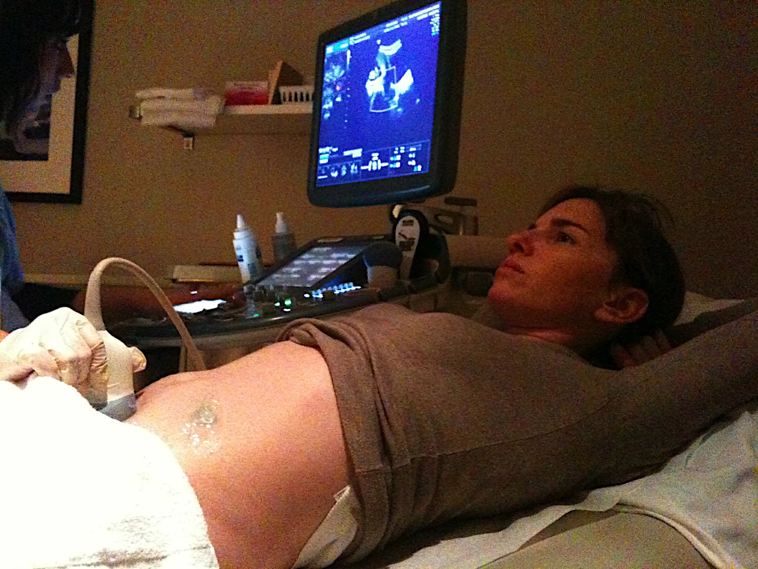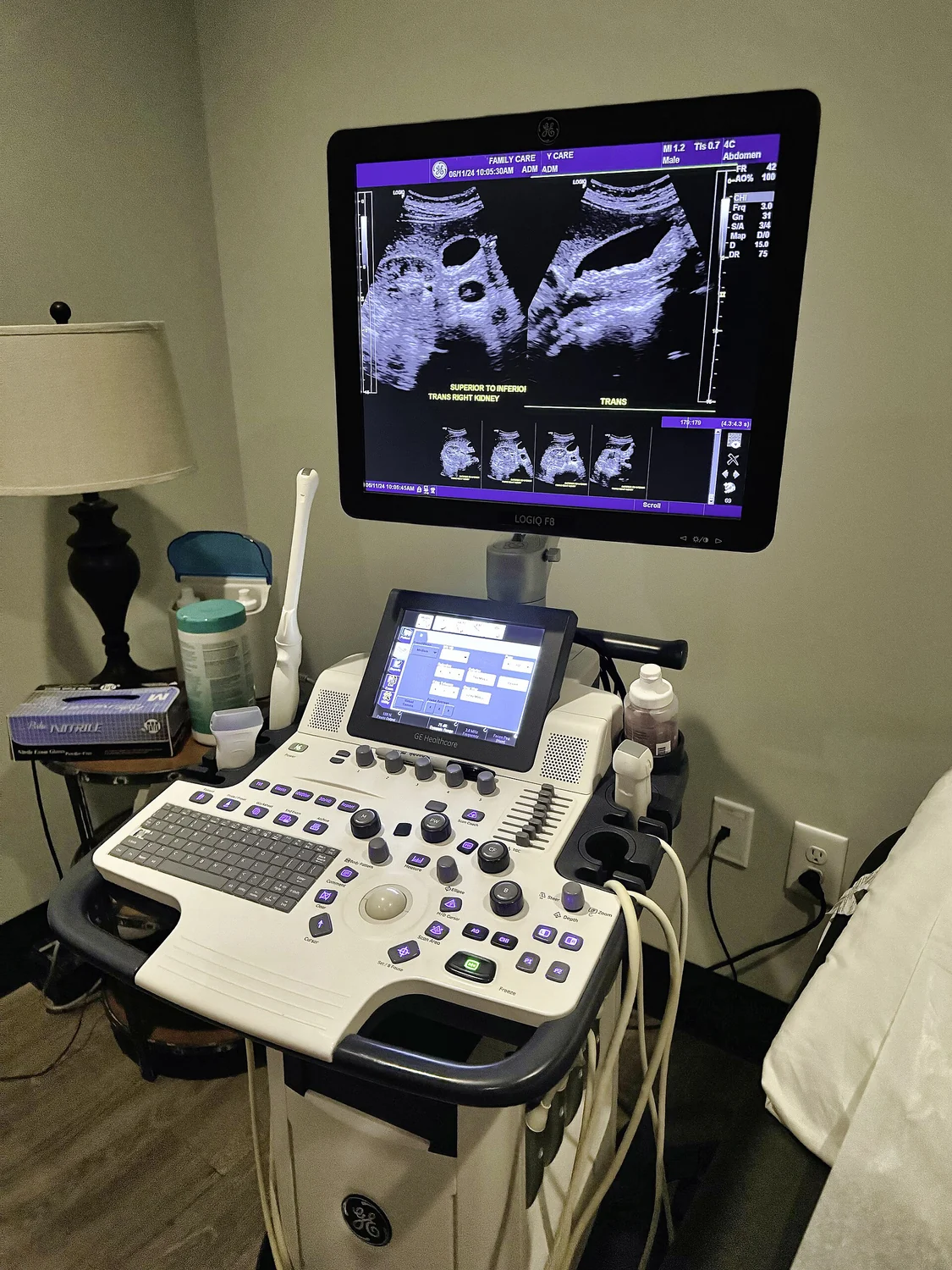
If ultrasound does not use x-rays, why does anyone talk about limiting its use? The answer lies in the difference between ionizing radiation and energy more broadly. Diagnostic ultrasound sends high-frequency sound waves into the body and listens for returning echoes. It does not carry the same ionizing-radiation exposure associated with x-ray imaging, but sound energy still interacts with tissue—and those interactions depend on how the examination is performed.
Sound makes an image from echoes
An ultrasound transducer emits sound waves above the range of human hearing and detects echoes returning from boundaries between tissues. The time for an echo to return helps estimate distance, while its strength contributes information about the reflecting structure [2]. A computer assembles these measurements into an image. Gel helps remove air between probe and skin, because air can block sound transmission.
The same physical process can show motion as well as structure. In Doppler ultrasound, changes in returning signals help represent blood movement and direction; a real-time display can reveal flow through a vessel [2]. The FDA notes that ultrasound images can show organs moving and blood flowing, unlike a still photograph [1].
Nonionizing does not mean no interaction
X-rays are ionizing radiation: they can alter atoms by removing electrons. Ultrasound is a mechanical wave traveling through tissue, not an x-ray beam, and it does not expose a patient to ionizing radiation [1]. That is why it can be useful when clinicians need imaging without that particular exposure, when medically appropriate.
But a sound wave carries energy. The FDA says ultrasound can slightly heat tissues and, in some circumstances, produce small gas pockets in body fluids or tissues, a phenomenon called cavitation [1]. These are physical effects, not proof that a standard diagnostic scan causes harm. They explain why “nonionizing” is a precise description of one kind of risk, not a synonym for “no biological effect.”
Imaging and treatment use different intensities
Diagnostic ultrasound is designed to gather information. Therapeutic ultrasound has a different purpose: its acoustic output can be used to heat, move, or destroy tissue, and focused high-intensity ultrasound is investigated or used in particular applications [2]. The distinction is like comparing a microphone listening to a sound with a speaker concentrating energy on a target: both involve waves, but their intended interaction differs.
The FDA advises that diagnostic ultrasound be used prudently by appropriately trained providers and that exposure be minimized while preserving diagnostic quality [1]. The amount and pattern of energy, the duration, and the tissue being examined all matter. A scan used to create an image should not be conflated with a treatment that deliberately modifies tissue.
Why controls and judgment remain useful
Ultrasound can be excellent for viewing many soft tissues, but it is not equally suited to every location. Air-filled regions and bone can block or complicate sound transmission, while fluid and soft-tissue boundaries often provide useful echoes [2]. A clinician selects imaging based on the question, anatomy, and information needed, not simply because one technology avoids ionizing radiation.
In pregnancy, ultrasound has important medical uses, while unnecessary prolonged exposure and operation by untrained users raise concern [1]. More generally, the technology’s safety record and its physical capacity to affect tissue can both be true. Sensible controls recognize that sound is a form of energy: the goal is to obtain useful information with appropriate exposure, not to treat nonionizing as an exemption from care.
Explore the physics: an explanatory diagram

Sources & further reading
- Ultrasound Imaging FDA
- Ultrasound NIBIB
Prepared for the independent PSI Center reading library. About edition dates and source checking.
This explainer describes scientific principles. It does not assess individual treatment choices.