Why does an ultrasound image show a bright boundary between two tissues when both are invisible to the eye? The answer begins with how sound travels through each material. A sound wave meeting a change in tissue may continue onward, but part of its energy can reflect back as an echo. The size of that reflection depends partly on acoustic impedance: a measure related to a material’s density and the speed of sound through it.
A material’s acoustic signature
Acoustic impedance describes how a material responds to a passing sound wave. In simple terms, it depends on the material’s density and the speed at which sound travels through it. When a wave reaches a boundary between materials with different impedances, the change can send some sound back toward its source. A larger mismatch generally produces a stronger reflection, while closer-matched materials allow more sound to pass across the boundary.
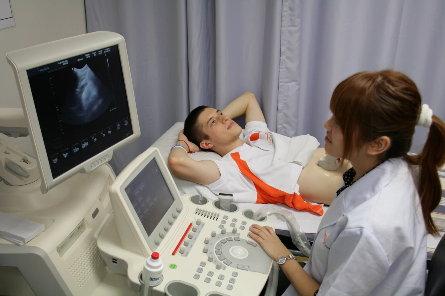
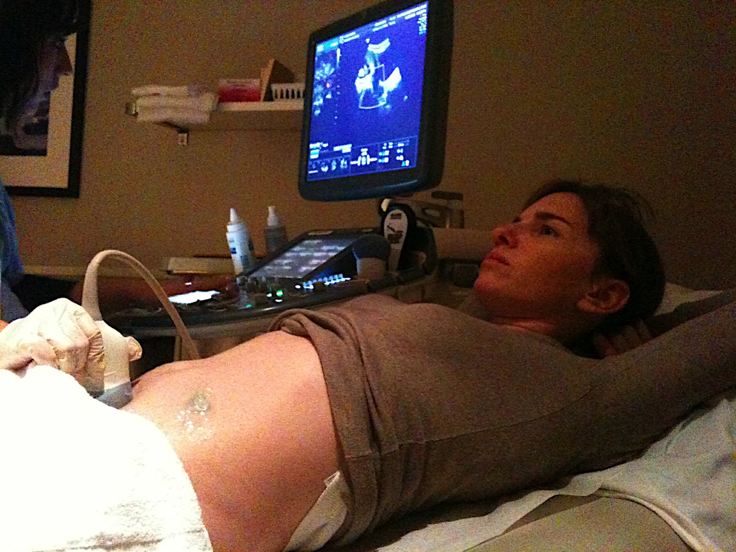
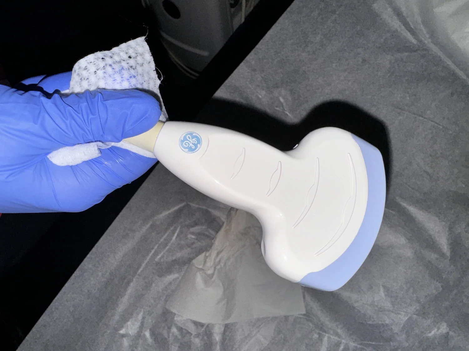
The boundary matters more than a tissue’s name alone. Imagine sound moving from soft tissue into a pocket of air: the transition is abrupt, and little sound may pass effectively across it. Ultrasound gel helps by filling small air gaps between the probe and skin, so the wave can enter the body instead of bouncing at the surface. [1]
Echoes become distances
An ultrasound transducer sends sound pulses into the body and detects returning echoes. The scanner measures how long each echo takes to return. Knowing the speed of sound in tissue, it estimates the distance to the reflecting boundary; many such measurements are assembled into an image. The brightness at a location reflects returning signal strength, which depends on the boundary and the path the sound took. [1]
This is why the image is not a direct photograph of every internal structure. It is a map built from echoes. A strong reflection can mark a sharp interface, while a weaker one can arise where neighboring tissues have more similar acoustic properties. Sound that is reflected or weakened before reaching deeper structures can limit what appears behind an interface. The image is information about how sound interacted with tissue, translated into a visual form.
The same physics, different biological job
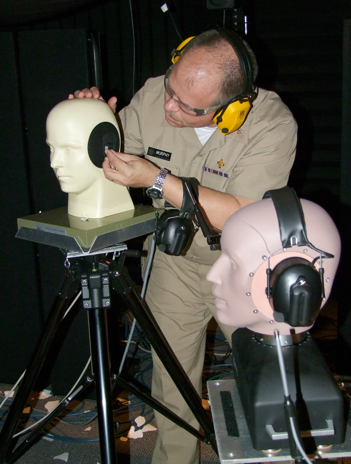
Sound also crosses boundaries in the inner ear, but there it is not being turned into a diagnostic image. Vibrations pass through the eardrum and tiny middle-ear bones to fluid in the cochlea. The fluid’s motion makes a traveling wave along the basilar membrane; hair cells move with it, bending tiny projections and triggering electrical signals that travel along the auditory nerve to the brain. [2]
This comparison highlights two different uses of mechanical waves. Ultrasound equipment sends pulses outward and analyzes echoes that return from tissue interfaces. Hearing depends on a chain that converts incoming vibrations into nerve signals. In both cases, sound must move through materials and set structures or fluids in motion, but the sensors and biological tasks are different. Acoustic impedance helps describe transmission and reflection; it does not by itself explain the full process of hearing or image formation.
Where an echo is useful—and where it is not
Diagnostic ultrasound is useful for many organs and for visualizing moving blood with Doppler methods, which can show flow direction and speed. But sound waves do not image all materials equally well. Ultrasound is not good for imaging bone or tissues containing air, because those interfaces interfere with the path into deeper regions. This is a physical limitation of sound transmission, not a failure of the scanner to create an image. [1]
The strength of an echo is also not a diagnosis by itself. Clinicians interpret patterns in context, using the anatomy, examination purpose and other information available. Ultrasound can produce anatomical images or functional maps, including measures of blood flow and tissue stiffness, but those are distinct techniques built on related physical measurements. A boundary’s echo tells the scanner that sound encountered a change; understanding what that change means requires interpretation.
Explore the physics: an explanatory diagram

Sources & further reading
- Ultrasound NIBIB
- How Do We Hear? NIDCD
Prepared for the independent PSI Center reading library. About edition dates and source checking.
This explainer describes scientific principles. It does not assess individual treatment choices.