If ultrasound is sound, why can’t we hear it? Its frequency is above the range human hearing can detect, but the basic physics remains familiar: it is a mechanical pressure wave moving through matter. A medical probe sends those waves into the body and listens for echoes, or, in therapeutic applications, concentrates energy to change tissue. Frequency helps distinguish ultrasound from audible sound; purpose and intensity distinguish one medical use from another.
Frequency beyond the ear
Sound is a traveling pattern of compressions and rarefactions—regions where material is slightly denser or less dense than its surroundings. Unlike light, it needs a material medium such as air, water, or tissue. Frequency counts how many cycles pass a point each second. Human hearing typically extends up to roughly 20 kilohertz; medical ultrasound uses frequencies above that threshold, commonly in the megahertz range [1].
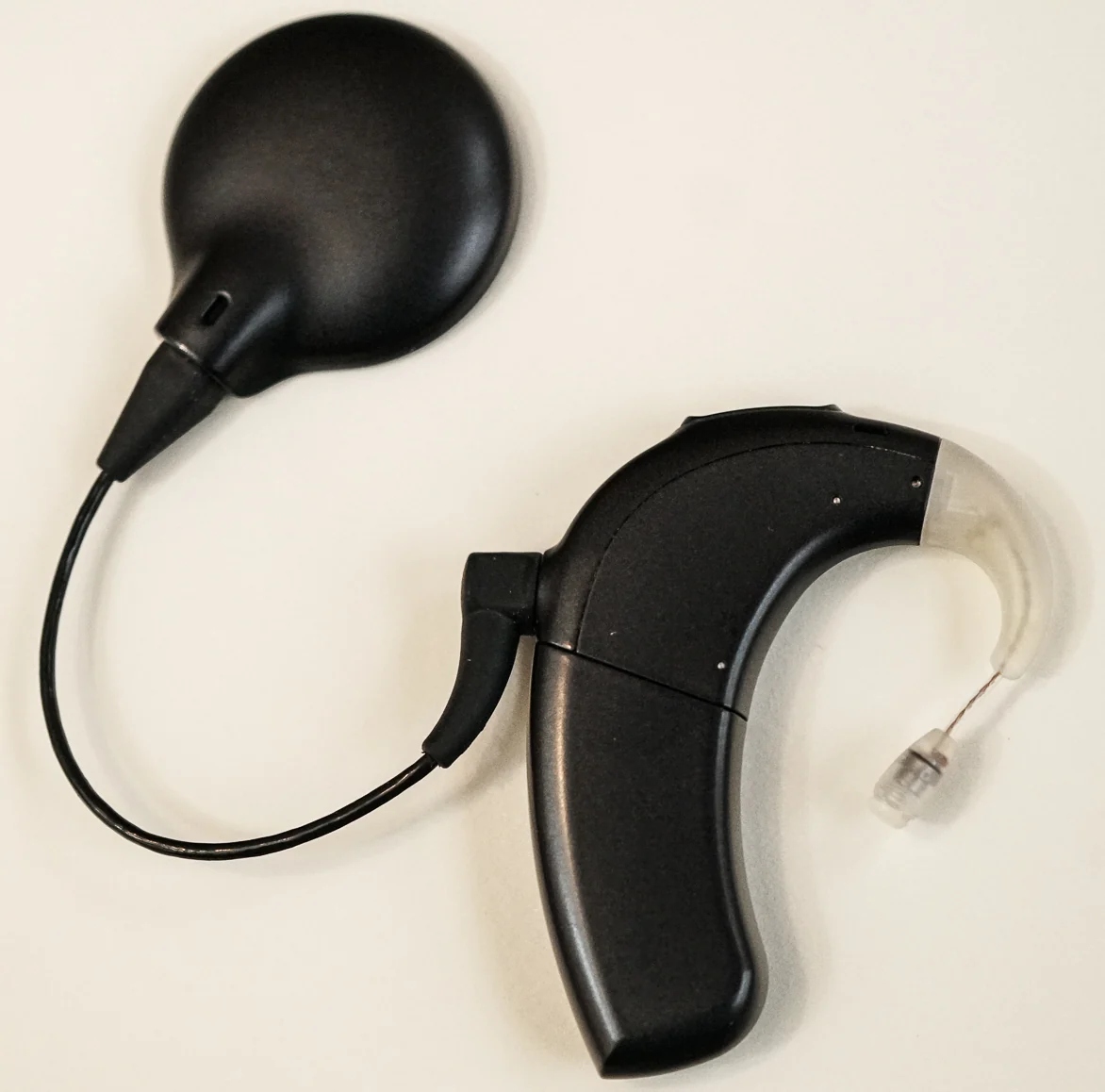
The ear converts sound vibrations into nerve signals through a sequence of mechanical steps. Vibrations move the eardrum and middle-ear bones, then ripple fluid in the cochlea, where hair cells turn motion into electrical signals [2]. Ultrasound waves can be sound without producing a conscious sound sensation because their frequency lies beyond the ear’s useful range.
Echoes become an image
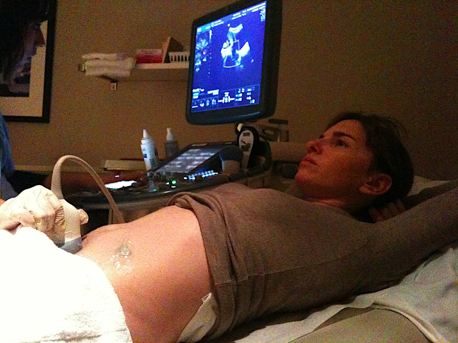
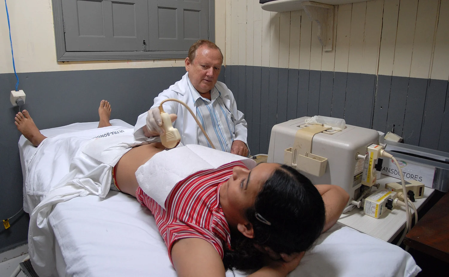
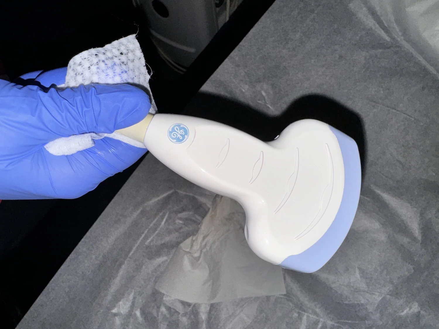
An ultrasound transducer both sends waves and detects returning echoes. Many probes use piezoelectric ceramic: applying an electric field makes it vibrate and produce sound; incoming sound makes it generate an electrical signal [1]. Tissue boundaries reflect part of the wave. By measuring the time for echoes to return and using the speed of sound, the scanner estimates how far away a boundary is and builds an image.
A smear of gel between probe and skin removes air pockets that would block waves from entering the body [1]. Think of calling into a cave: an echo arriving sooner usually comes from a nearer surface. The analogy is useful, though the body has many boundaries and the scanner turns thousands of echoes into a structured picture. Ultrasound works well for many soft tissues but struggles when sound meets bone or air-filled regions [1].
Imaging and therapy are distinct jobs
Diagnostic ultrasound aims to form information about anatomy or function. Doppler techniques use changes in echoes to display blood flow, while elastography estimates relative tissue stiffness [1]. Such modes add information to an anatomical picture; they do not mean the machine directly sees every biological process or automatically diagnoses disease.
Therapeutic ultrasound uses acoustic energy to modify or destroy tissue rather than produce an image. Focused high-intensity beams can heat or ablate selected tissue; other approaches are being investigated for tasks such as breaking clots or delivering drugs [1]. The distinction is like the difference between a flashlight used to inspect an object and a magnifying glass concentrating sunlight: related waves, different energy delivery and intent.
Safety follows the settings
Diagnostic ultrasound is generally regarded as safe and does not use ionizing radiation, but under specific settings and conditions sound can produce biological effects in tissue [1]. That is why regulatory limits and clinical use matter. The absence of x-rays does not make every possible ultrasound exposure identical or consequence-free.
The same term also covers technologies at different stages of use. Some therapeutic applications have approved medical uses, while other possibilities remain under investigation [1]. For example, researchers have studied wearable ultrasound patches and new focused-energy techniques, but an early study or proposed application is not evidence that every device is proven for routine care. The practical question is always what the waves are being used to measure or change, and under what controlled conditions.
Explore the physics: an explanatory diagram

Sources & further reading
- Ultrasound NIBIB
- How Do We Hear? NIDCD
Prepared for the independent PSI Center reading library. About edition dates and source checking.
This explainer describes scientific principles. It does not assess individual treatment choices.