If ultrasound is not an x-ray, what exactly enters the body during a scan? A probe sends high-frequency sound waves through gel and tissue; returning echoes help form an image. That process avoids ionizing radiation, but it is not energy-free. Sound can transfer energy to tissue, so “prudent exposure” means using ultrasound for a clear medical purpose and limiting unnecessary time or intensity while retaining the information needed for a useful examination.
Echoes turn sound into an image
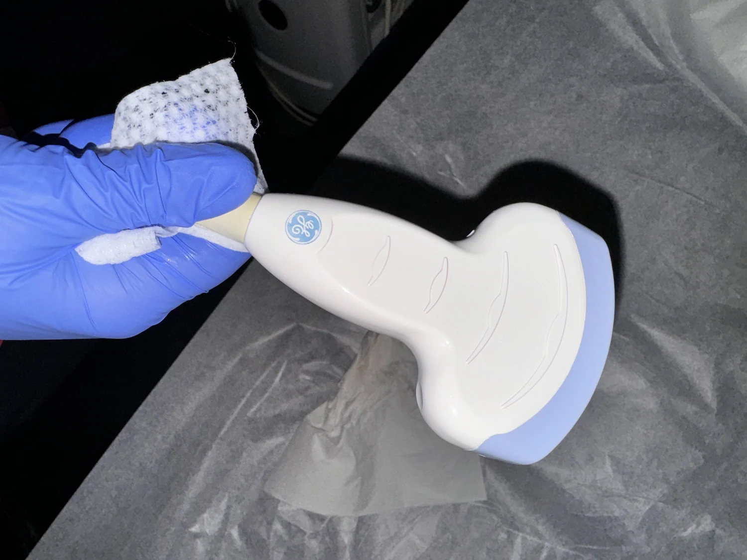
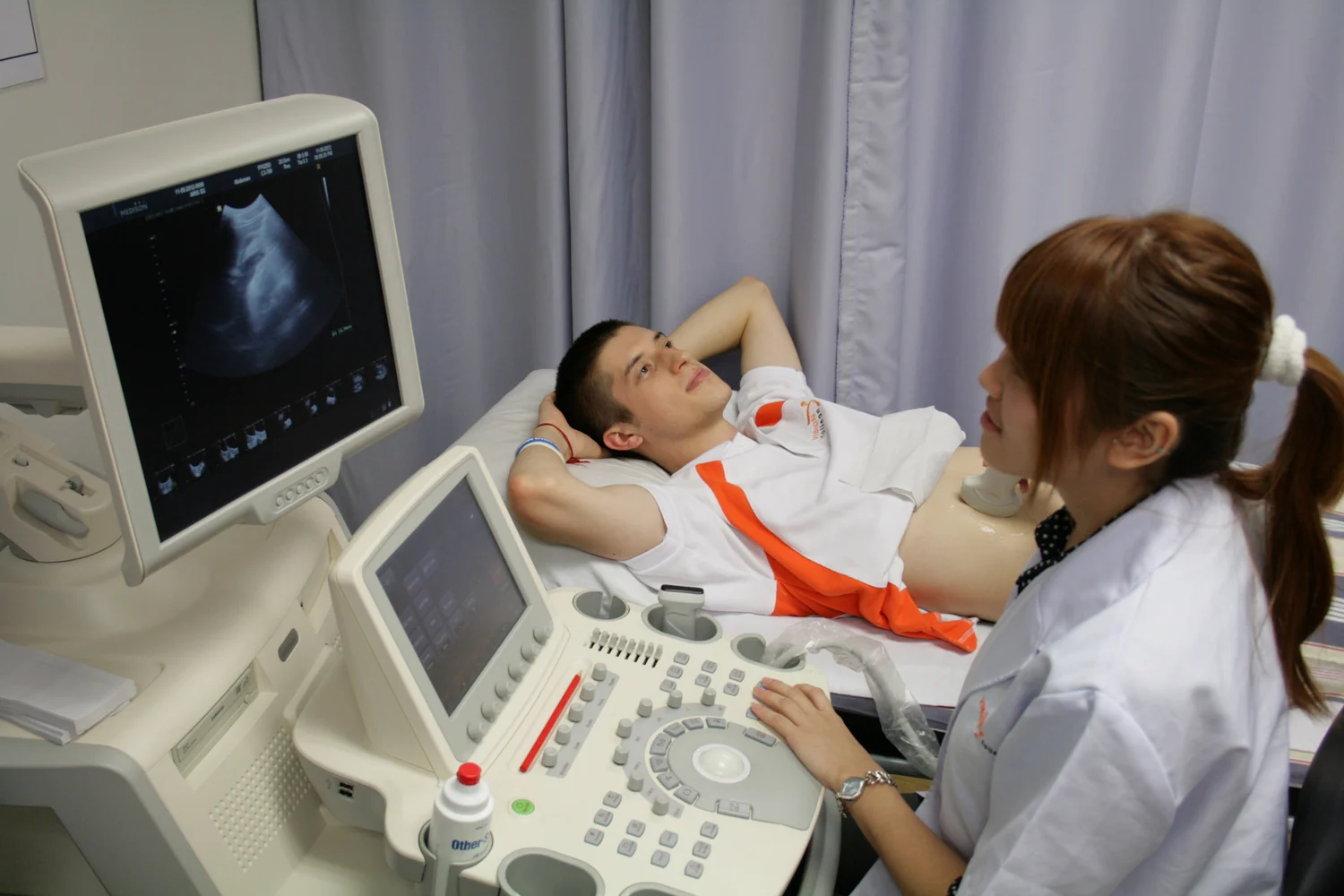
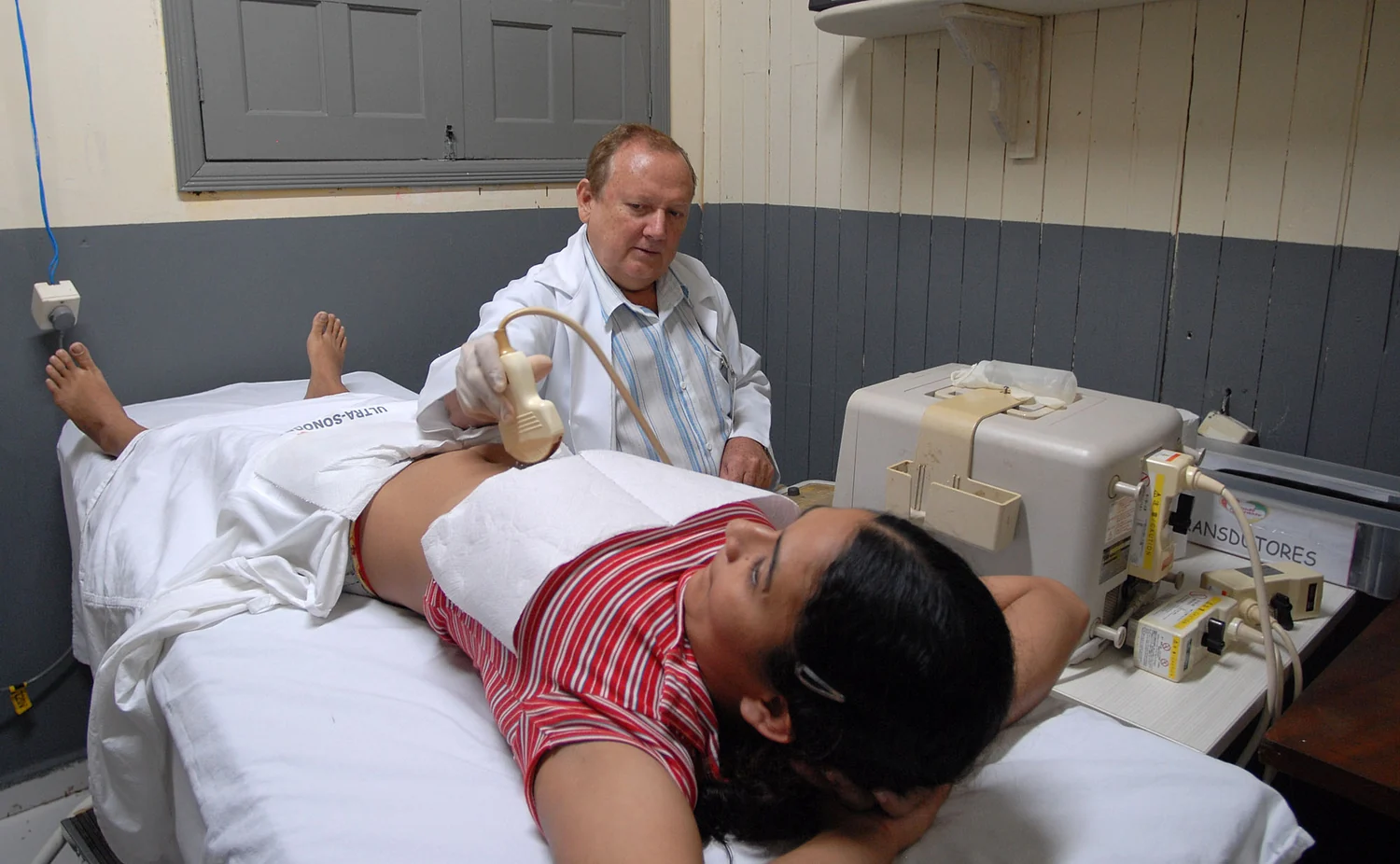
Ultrasound imaging uses sound waves at frequencies higher than people can hear. A transducer, or probe, sends pulses into the body and receives echoes reflected by boundaries between structures. The echo’s strength and the time it takes to return help a system construct an image. Because scans can be captured in real time, clinicians can observe movement and blood flow as well as still anatomy [1].
A simple mental picture is shouting across a canyon and estimating the wall’s distance from the returning echo. A medical scanner does something more sophisticated: it emits many short pulses, detects their returns, and combines information from different directions. The probe’s gel layer helps sound pass from the probe into the body rather than being trapped by air at the skin surface. The image depends on reflected sound, not on a beam of x-rays passing through the body [1].
Non-ionizing does not mean no interaction
X-rays are ionizing radiation: their energy can potentially damage DNA. Diagnostic ultrasound instead uses mechanical sound waves and is not associated with ionizing radiation exposure [2] [1]. That distinction matters, but it does not mean ultrasound has no physical effects. The waves can heat tissue slightly, and under some conditions can produce cavitation, small pockets of gas in fluids or tissue. The long-term consequences of these effects are not fully known [1].
The word “mechanical” is useful here. A sound wave is a traveling pressure disturbance: neighboring particles move back and forth, passing energy along without the wave being an x-ray. If the energy is absorbed, tissue may warm; under some conditions, pressure changes may affect gas bubbles. These mechanisms do not establish that a diagnostic examination causes harm. They explain why a technology with a strong safety record is still used with attention to exposure and clinical purpose [1].
Prudent use connects purpose to exposure
The FDA describes ultrasound as generally considered safe when used prudently by appropriately trained health care providers. Prudent use includes considering how much exposure is needed to obtain diagnostic-quality information and minimizing exposure while maintaining that quality. This resembles the broader idea of optimization in medical imaging: settings and procedure should match the question, rather than adding exposure without a reason [1].
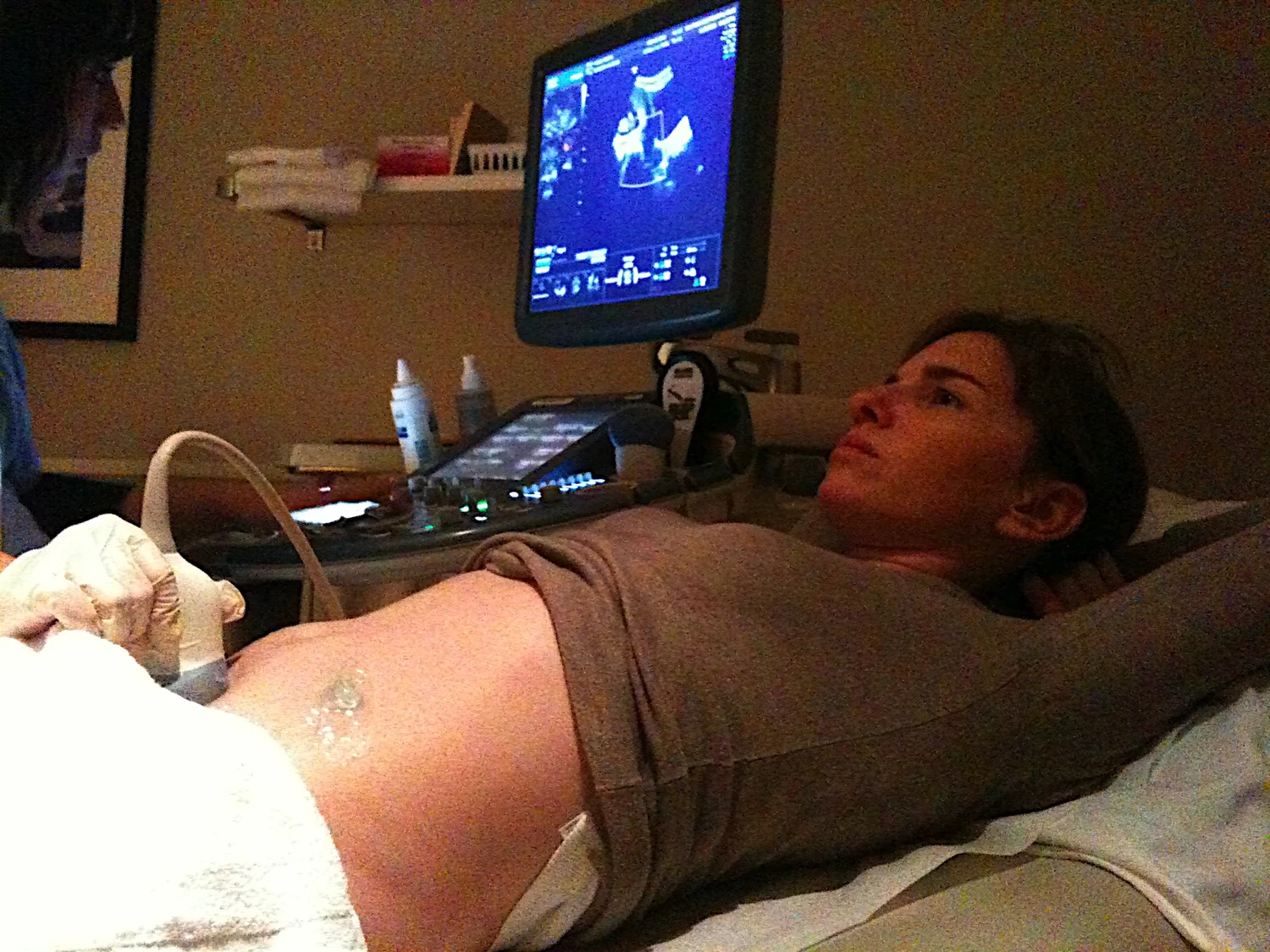
Pregnancy makes the principle especially visible. Ultrasound is widely used to assess and monitor fetal and maternal health, but prolonged or unnecessary exposure may increase risk, and untrained operation can produce unsafe exposure or misleading interpretations. The FDA discourages using ultrasound solely to create keepsake videos when extra exposure is required. Images captured as part of a medically indicated exam do not require an added scan just for keepsake purposes [1].
Different imaging methods answer different questions
A comparison with x-ray imaging helps avoid a false choice between “safe” and “dangerous.” X-rays can be valuable when a clinical question calls for them; their ionizing energy carries a small potential cancer risk that depends on exposure and other factors. Clinicians weigh benefits against risks and seek to avoid unnecessary exposure [2]. Ultrasound may be suitable for some questions, but it is not automatically a substitute for every other modality. Its value depends on the body region and information sought.
For example, a real-time view of blood moving through a vessel is a strength of Doppler ultrasound, while other tests may better answer different questions. Prudent practice is therefore not a command to avoid ultrasound or to use it without limit. It is a way of matching the scan to a medical purpose, using trained operators and an exposure sufficient for the task. That balance preserves the practical benefit of seeing internal motion while respecting that sound energy interacts with tissue [1] [2].
Explore the physics: an explanatory diagram

Sources & further reading
Prepared for the independent PSI Center reading library. About edition dates and source checking.
This explainer describes scientific principles. It does not assess individual treatment choices.