How does a machine turn a faint echo into a point on a picture? It measures how long sound takes to travel into the body and back. If the sound speed is known, that round-trip time gives an estimate of the reflector’s distance. Repeating the measurement across many pulses builds a map of tissue boundaries. The basic trick resembles listening for an echo in a canyon, but the scanner does the timing and placement at scales far too small for the ear.
First, make a pulse and catch its return
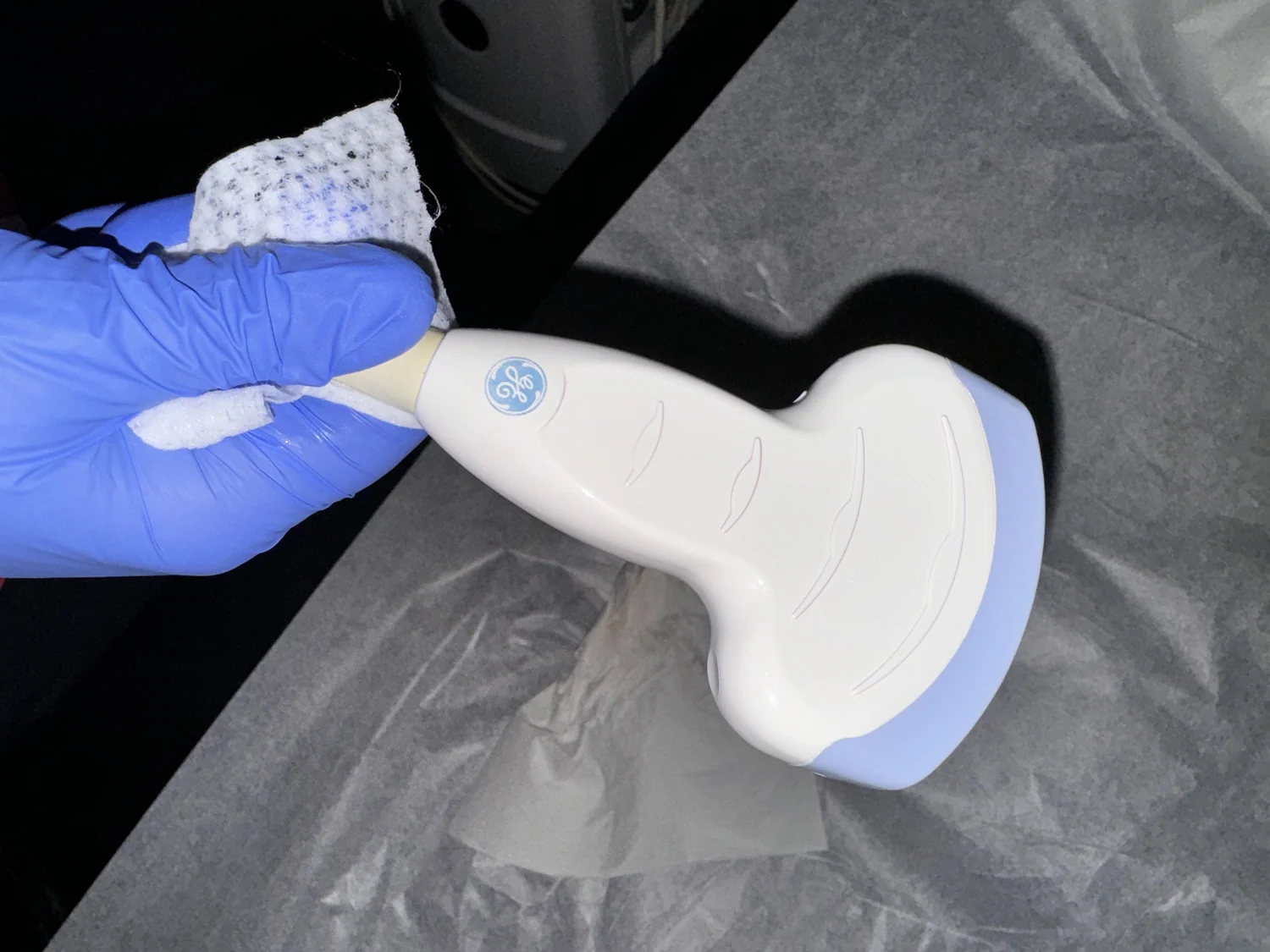
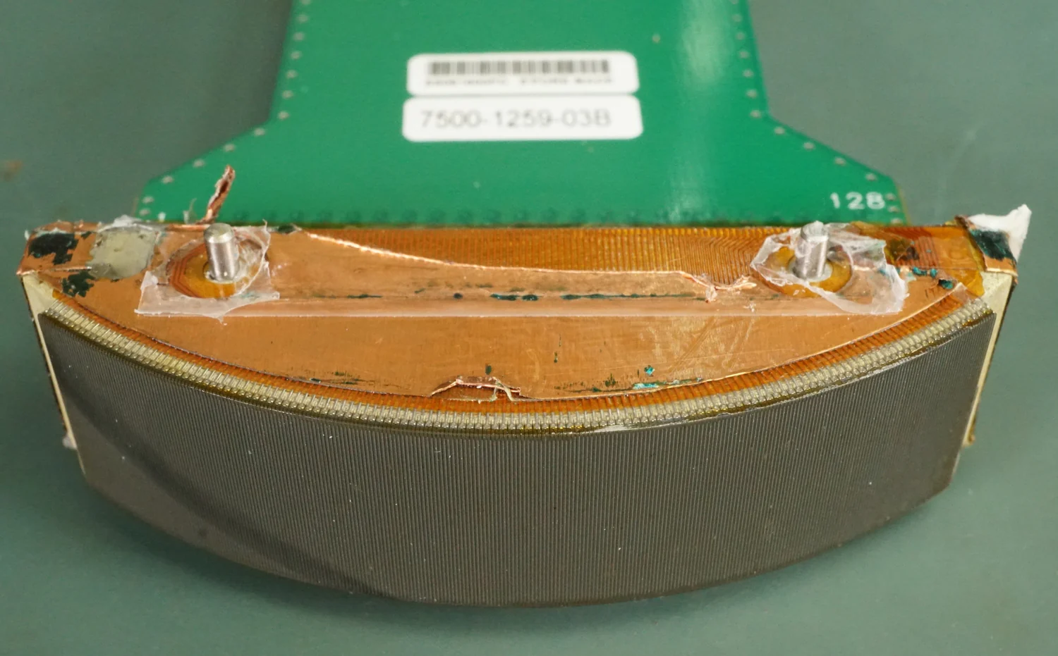
An ultrasound probe, also called a transducer, sends sound waves above the range of human hearing into the body. Many probes use piezoelectric crystals: applying an electric field makes them vibrate and produce sound, while returning sound makes them generate an electrical signal. The same probe can therefore transmit a pulse and receive its echoes [1].
Sound reflects when it meets a boundary between materials with different acoustic properties, such as fluid and soft tissue or tissue and bone. Some sound continues onward, and some returns to the probe. The scanner records the time between sending a pulse and detecting an echo. That elapsed time is the key measurement; the machine does not directly sense depth as a camera might sense a surface [1].
The round trip is twice the distance
Suppose a pulse takes a certain time to travel from the probe to a boundary and back. If the speed of sound through the tissue is represented by a known value, multiplying speed by total travel time gives the path length. Because the pulse travelled out and back, the one-way depth is half that total. In compact form: depth equals sound speed multiplied by echo time, divided by two.
The factor of two is easy to miss. Imagine standing a known distance from a wall and timing a shout’s echo. The sound covers the distance to the wall and the same distance back to you. Ultrasound uses the same logic, but at microscopic scales and with pulses that return in fractions of a second. The speed used is an estimate for the tissues being scanned, so the resulting location is an estimated depth rather than a ruler reading.
Many echoes become an image
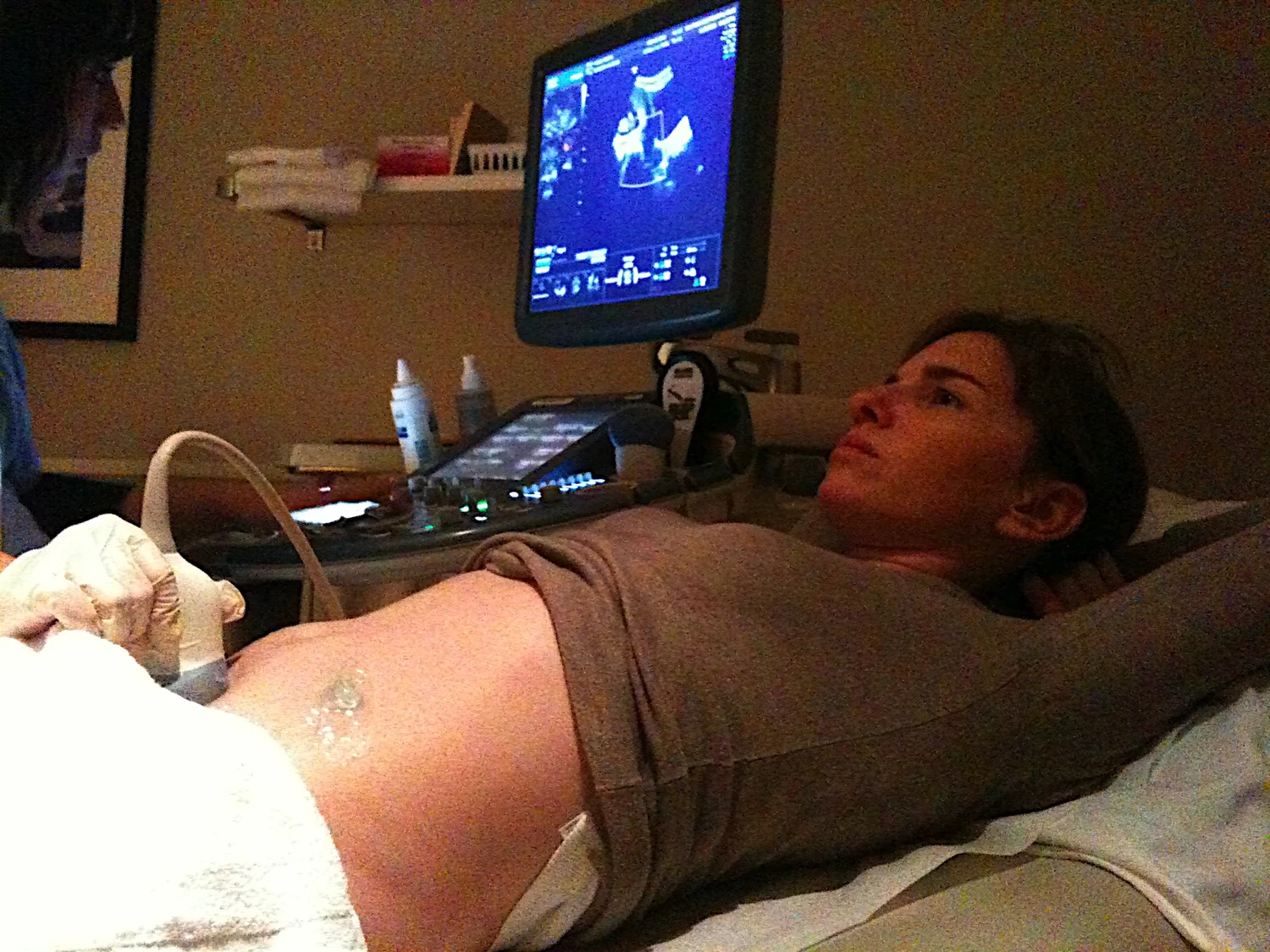
One echo marks a reflecting boundary along the direction of a pulse. A scanner sends pulses repeatedly, often steering or moving the beam across the region, and records the return times and strengths. The time information places echoes at depths; their strengths contribute to image brightness. Combining these measurements across positions creates a two-dimensional representation of structures, with additional methods producing three-dimensional or moving views [1].
The mental experiment can be extended: point the echo timer in many directions rather than toward one wall. Each direction produces a line of distances. Assemble the lines and a pattern of boundaries emerges. Diagnostic ultrasound uses this kind of return-signal mapping to show internal organs and other structures. Functional approaches can add information such as blood-flow movement or relative tissue stiffness to anatomical images [1].
Why contact and assumptions matter
Air between the probe and skin would reflect much of the sound before it entered the body. Coupling gel fills tiny gaps and helps sound pass from probe into skin [1]. Once in the body, echoes can be weak or blocked by structures that reflect or absorb sound strongly. Ultrasound is therefore useful for many soft tissues but is not generally good for imaging bone or air-filled regions such as the lungs [1].
The depth calculation also assumes a sound speed for the path, even though real bodies contain layers with different properties. A mismatch can shift where an echo appears in the image. Computational modeling offers a broader way to understand such measurements: models use mathematics and physics to represent systems, simulate outputs, and examine how changed assumptions affect predictions [2]. The image is a carefully constructed interpretation of measured echoes, not a literal photograph of the inside of the body.
Explore the physics: an explanatory diagram

Sources & further reading
- Ultrasound NIBIB
- Computational Modeling NIBIB
Prepared for the independent PSI Center reading library. About edition dates and source checking.
This explainer describes scientific principles. It does not assess individual treatment choices.