Why can one sound pulse draw a picture while another is used to break a kidney stone? Both involve pressure waves traveling through material, but their shapes, intensities, and purposes differ. Diagnostic ultrasound sends pulses and reads returning echoes. Shock wave lithotripsy concentrates mechanical effects on a stone so fragments can pass through the urinary tract. Understanding the distinction starts with how an ordinary echo becomes an image. [2]
An echo is a timed reflection
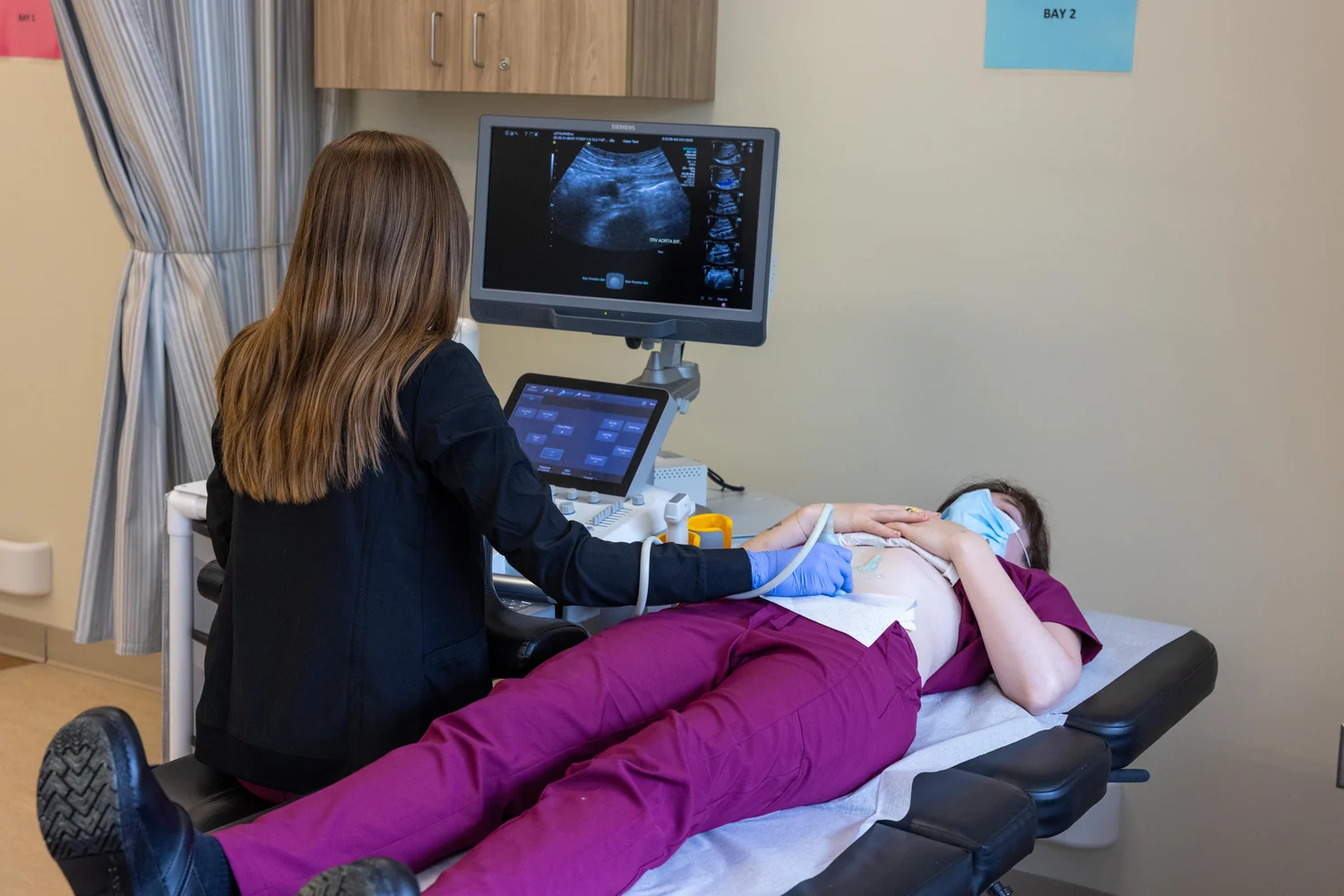
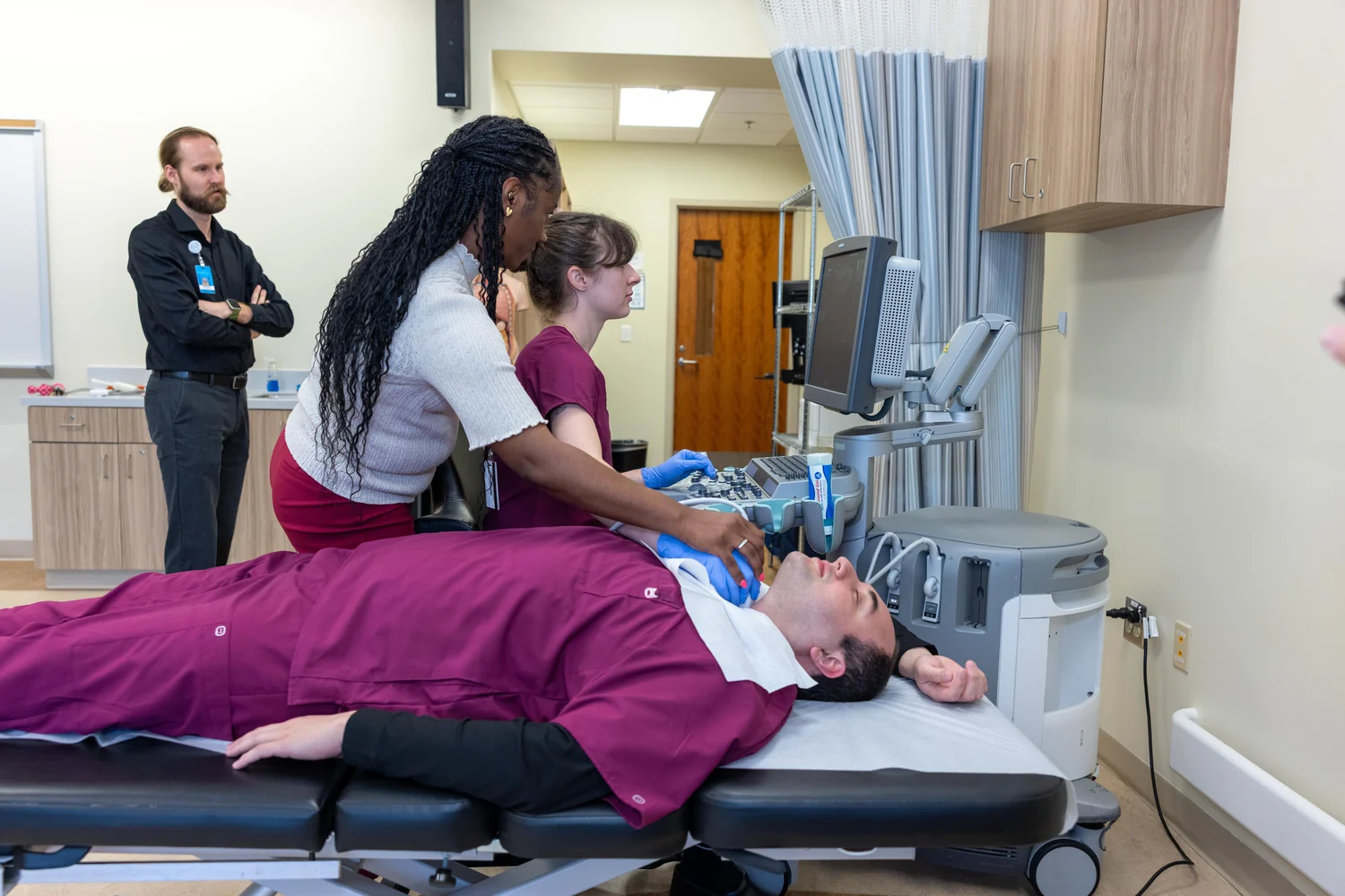
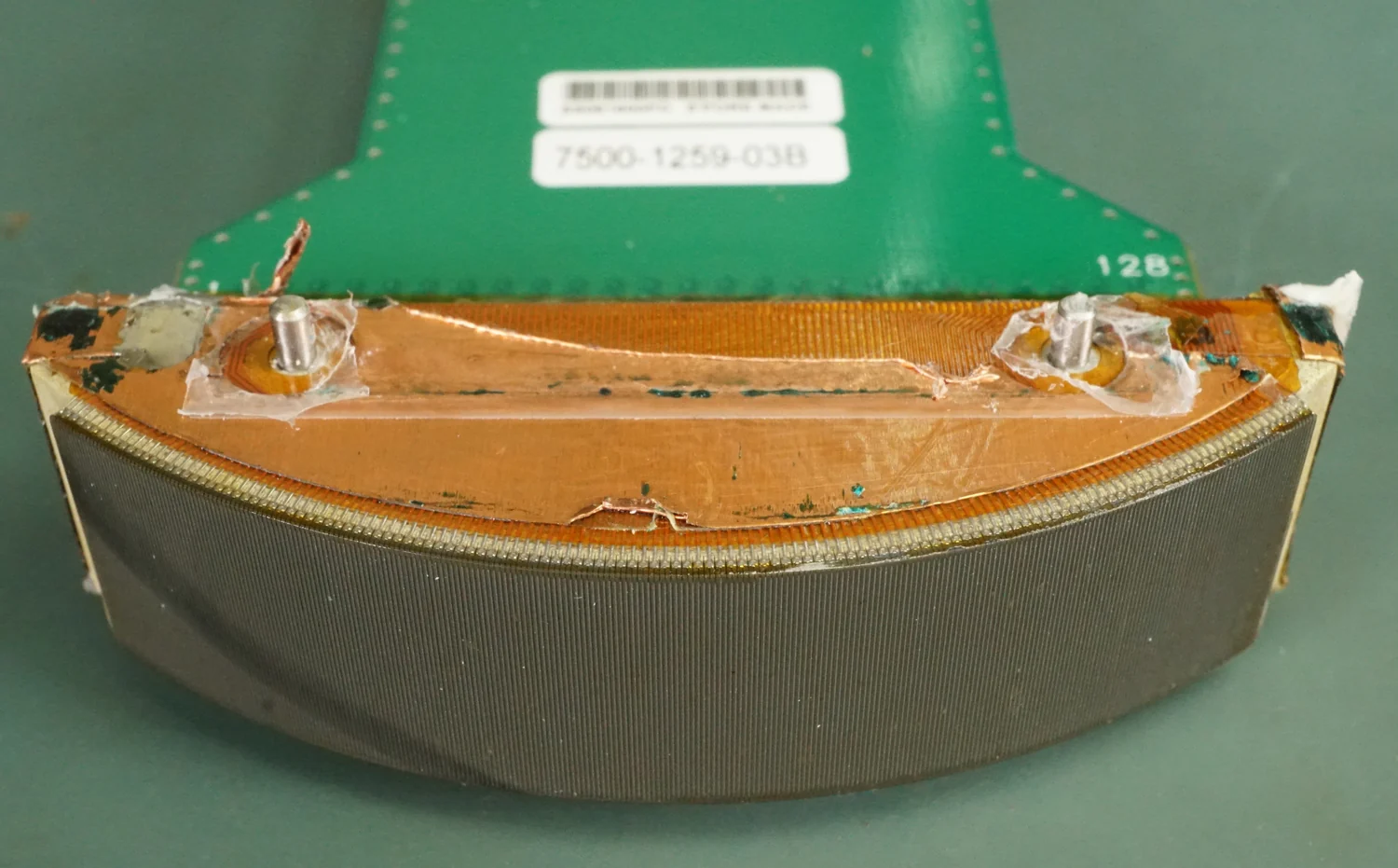
An ultrasound transducer both emits sound and detects echoes. Its piezoelectric elements convert electrical energy into a sound pulse, then convert returning sound into an electrical signal. Boundaries between tissues reflect part of the pulse; a scanner times each echo and uses the speed of sound to estimate the distance to that boundary. Repeated across directions, these measurements create images of internal structures. [2]
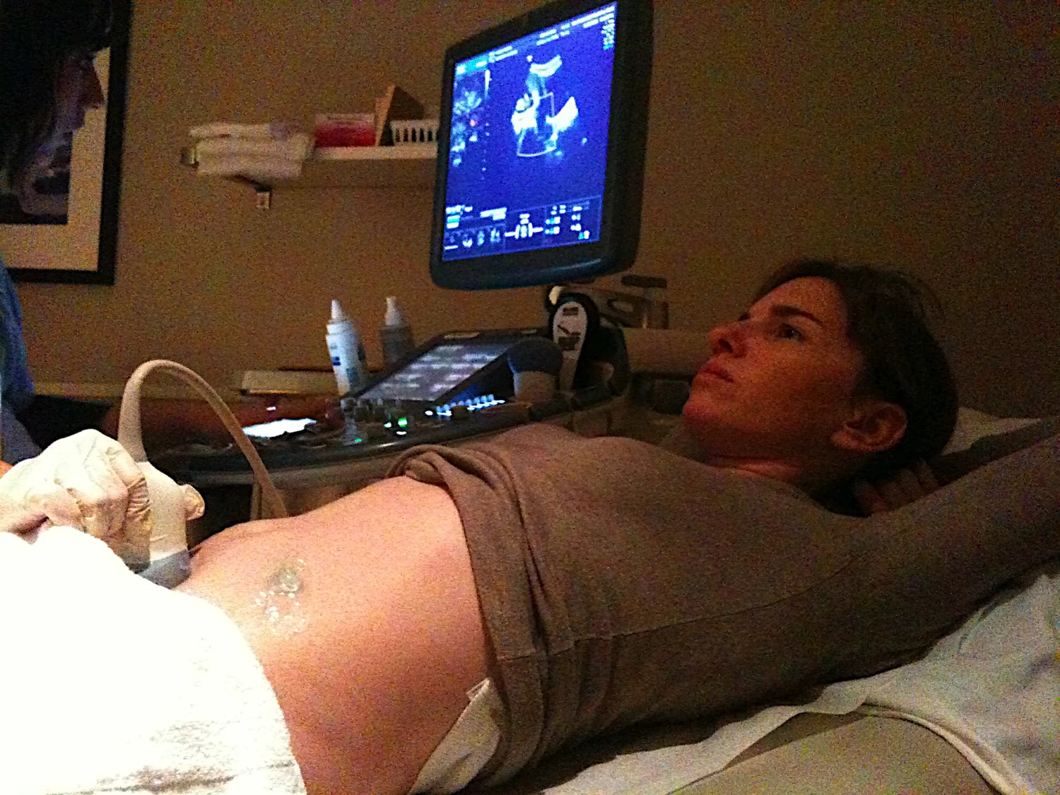
A useful mental picture is calling into a cave. The delay before the echo returns hints at the distance to a wall, while the echo’s strength carries information about the reflecting boundary. In the body, the scanner sends many such pulses and organizes their returns into a two-dimensional view. Gel helps by preventing air pockets between probe and skin, since air can block sound transmission. [2]
Imaging asks for information
Diagnostic ultrasound is designed to obtain information while imaging. A probe may show anatomy, or functional features such as blood movement and tissue stiffness can be combined with anatomy into information maps. In Doppler imaging, for example, reflected sound from moving blood shifts in frequency, allowing flow direction and speed to be displayed. The diagnostic task depends on interpreting echoes rather than mechanically disrupting a target. [2]
Sound does not travel equally well through every body region. Ultrasound is not good for imaging bone or air-filled tissue, though under some conditions it can image bones in fetuses or infants and fluid-associated regions of the lungs. These limits arise from how waves reflect and propagate at boundaries. They also underline that an ultrasound image is a product of both the tissue and the sound path. [2]
A shock wave delivers a mechanical effect
Shock wave lithotripsy is a treatment for kidney stones: a clinician uses shock waves to break a stone into smaller pieces that can then pass through the urinary tract. The goal is not to map a boundary from an echo. It is to direct acoustic energy so that the stone experiences mechanical stresses sufficient to fragment it. NIDDK lists the procedure among options for stone removal, with suitability depending on factors including stone size and location. [1]
A shock wave has a sharp pressure rise and a strong peak, unlike the comparatively low-intensity pulses used to form diagnostic pictures. Focusing matters: energy is concentrated at a selected location, while the therapeutic setup aims to limit effects outside the target. The word “sound” covers both processes, but does not imply they are interchangeable. One measures reflections; the other uses pressure to alter material.
Purpose determines the acoustic design
Therapeutic ultrasound is a broad category, not a synonym for lithotripsy. Some therapeutic systems heat or ablate tissue, while others move tissue, dissolve clots, or deliver drugs. High-intensity focused ultrasound is investigated or used in specific contexts to modify or destroy targeted tissue. These are distinct approaches because the desired physical interaction differs, even though each uses sound above human hearing. [2]
After lithotripsy, the fragments still have to leave the body through the urinary tract; breaking a stone is not the same as removing every fragment instantly. NIDDK notes that larger or obstructing stones and severe pain may require urgent treatment, and that clinicians choose among procedures based on the situation. The physics explains how a wave can fragment a target, while clinical decisions depend on the individual stone and patient. [1]
Sources & further reading
- Treatment for Kidney Stones NIDDK
- Ultrasound NIBIB
Prepared for the independent PSI Center reading library. About edition dates and source checking.
This explainer describes scientific principles. It does not assess individual treatment choices.