Why does one patch on an ultrasound screen look bright while the neighboring region appears dark? The answer begins with sound reflecting at boundaries inside the body, then continues through choices made by the scanner that turns echoes into pixels. A bright echo can reveal a physical difference, but it does not name the cause of that difference. The image is evidence to interpret, not a diagnosis by itself.
The returning pulse
An ultrasound transducer sends pulses of sound into the body. The frequencies are above human hearing, and medical probes commonly operate in the megahertz range. When a pulse meets a boundary between materials with different acoustic properties, some sound reflects back toward the probe. Fluid next to soft tissue, for example, can produce an echo because the wave encounters a change in its surroundings [1].
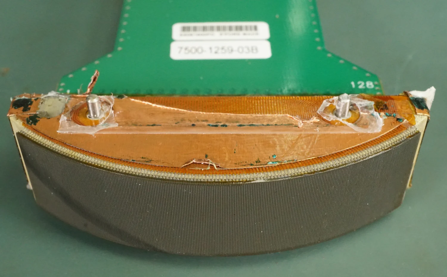
The same probe listens for returning echoes. Many probes contain piezoelectric materials: crystals that change shape under an electric field and, in reverse, produce electrical signals when pressure from an echo makes them vibrate. The scanner measures how long each echo takes to return. Given the assumed speed of sound through tissue, it estimates the boundary’s depth and places a mark at that location. Repeating this across many directions builds an image.
Brightness is a measurement choice
In a typical two-dimensional image, stronger returning echoes are displayed as brighter pixels and weaker echoes as darker ones. This is a useful convention, not a direct photograph of tissue. The display compresses and maps measured signals into a limited range of shades. Gain, focus, probe angle, and other settings can change the appearance. A region that looks conspicuously bright is therefore a prompt to ask what reflected sound and how the scanner rendered it.
A simple mental experiment makes the distinction clear. Imagine shining a flashlight across a rippled pond: some angles send a strong glint toward your eyes, while others send light elsewhere. The water has not changed, but your view has. Sound reflections also depend on geometry. A boundary tilted away from the probe may return less energy, while a change in tissue composition may return more. One bright patch cannot, by itself, distinguish these possibilities.
Anatomy, function, and context
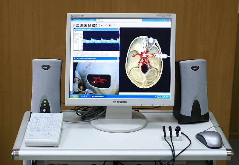
Diagnostic ultrasound can show anatomy, such as the shape of an organ, or functional information such as blood movement. Doppler techniques use changes in returning sound to estimate blood-flow direction and speed; color maps can display that flow over anatomy. Elastography uses tissue motion or deformation to estimate relative stiffness. These modes add different measurements, yet each still depends on how a signal was acquired and processed [1].
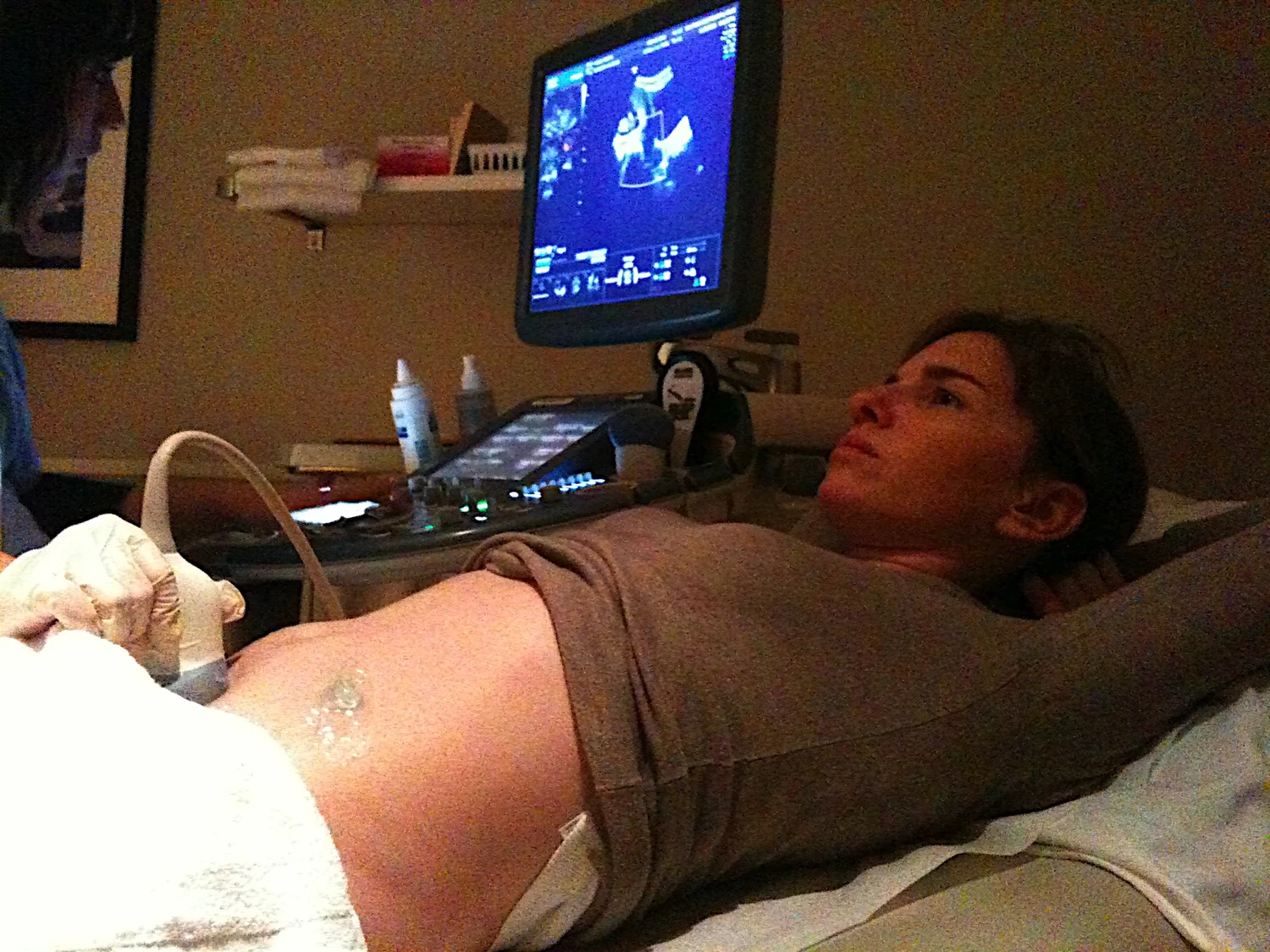
The body also creates blind spots. Air and bone impede ultrasound, so lungs and adult bones are generally difficult to image, although particular conditions can allow views of the lung lining or certain bones. Gel helps by eliminating air pockets between skin and probe, which would otherwise block transmission. Probe position, the tissue along the beam, and the question being examined all shape what can be seen. Medical devices more broadly may monitor, modulate, or supplement bodily functions, but their measurements serve defined purposes rather than automatically explaining every symptom [2].
From signal to clinical judgment
A clinician combines the image with the reason for the examination, other findings, and the limits of the technique. A bright or dark region could reflect a boundary, a material property, the angle of observation, or a setting. In ultrasound-guided biopsy, for instance, real-time images help show where a needle is relative to a target. The image guides a physical procedure; it does not replace examination of the sampled tissue when that is needed [1].
Diagnostic ultrasound does not use ionizing x-rays, and it is generally regarded as safe. Still, sound energy can produce biological effects under some conditions, which is why diagnostic devices operate within accepted limits and casual nonmedical scanning is discouraged [1]. The practical lesson is not to distrust a striking image, but to interpret it at the right level: it records echoes under particular conditions. A diagnosis requires connecting that record to anatomy, function, and clinical context.
Sources & further reading
- Ultrasound NIBIB
- Medical Device Technologies NIBIB
Prepared for the independent PSI Center reading library. About edition dates and source checking.
This explainer describes scientific principles. It does not assess individual treatment choices.