How can sound waves heat a small region deep inside the body while the skin remains uncut? Ultrasound can carry energy through tissue and be focused so that waves converge at a target. There, absorbed energy can raise temperature enough to alter or destroy tissue. The same word, ultrasound, also names familiar diagnostic imaging, but the goal and intensity differ: one method listens to echoes, the other deliberately deposits energy.
From echoes to energy
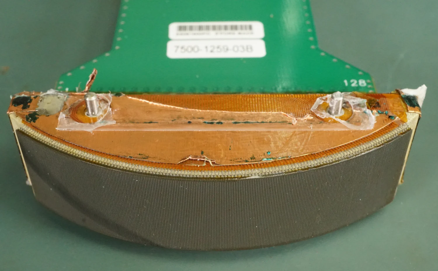
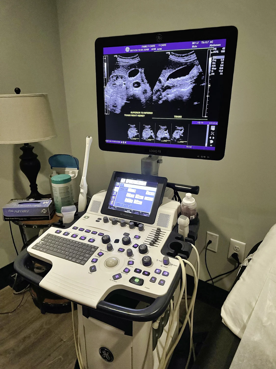
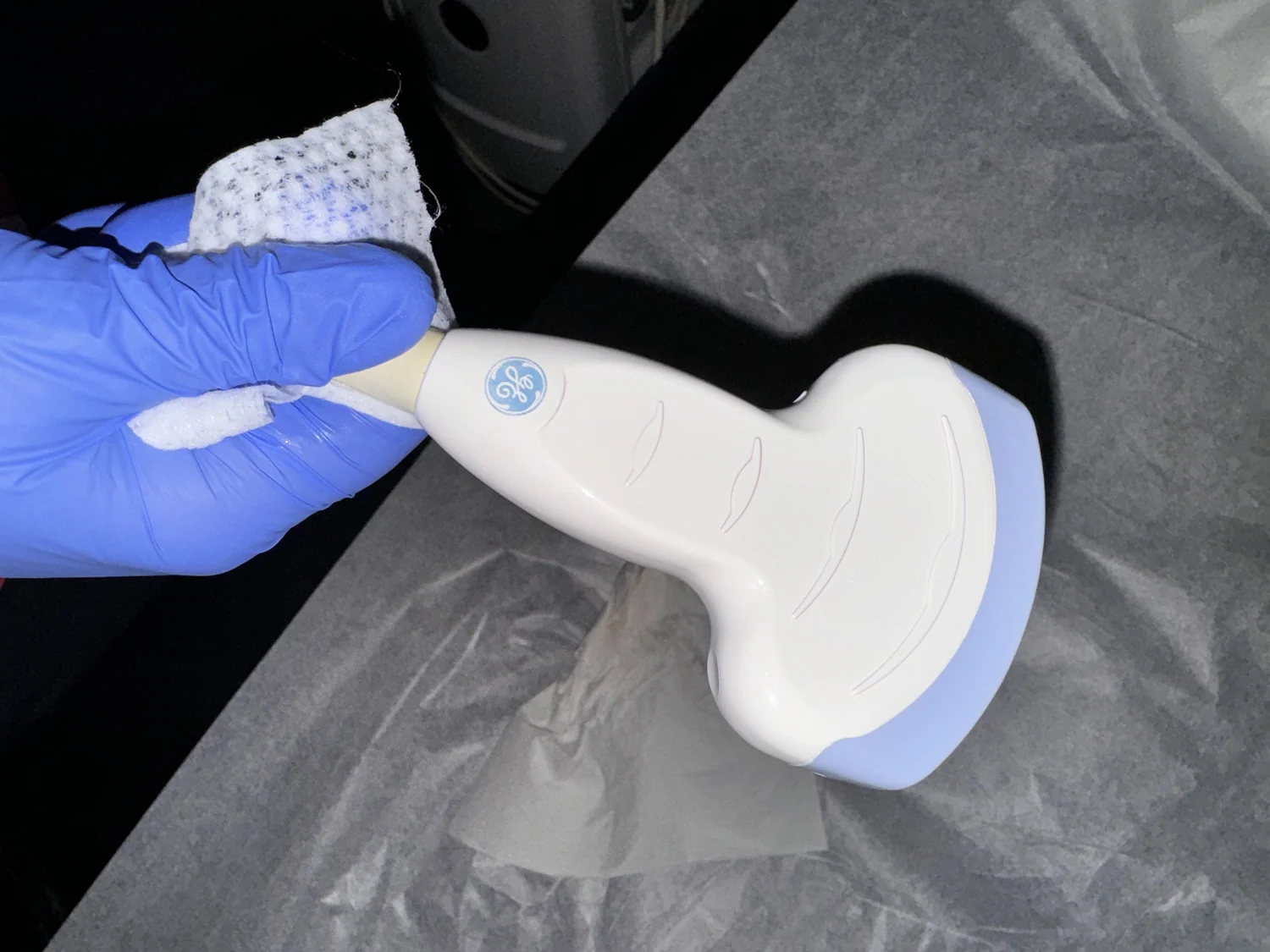
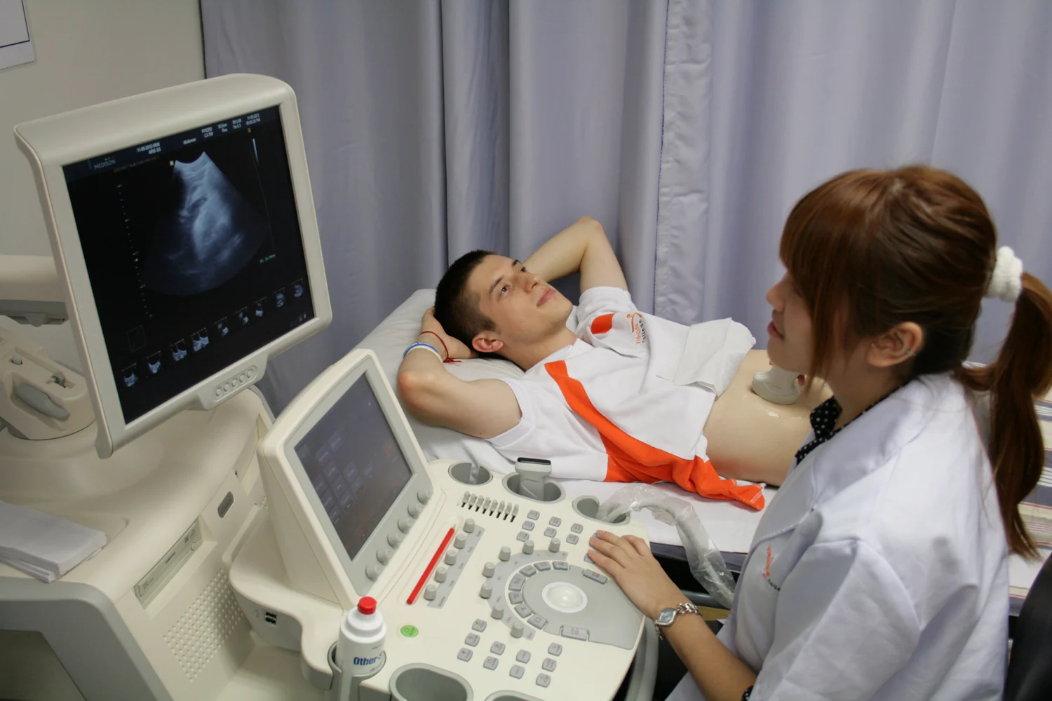
In diagnostic ultrasound, a transducer sends sound into the body and detects echoes returning from boundaries between tissues. Their timing helps the scanner estimate depth and build an image [1]. A therapeutic beam is designed for interaction rather than picture-making. At high intensity, sound energy can heat, move, or damage tissue; high-intensity focused ultrasound (HIFU) concentrates the beam at a selected location [1]. The difference is not simply that one uses sound and the other does not. It is how energy is delivered and what the procedure is meant to accomplish.
A useful analogy is sunlight through a magnifying glass: energy spread over a broad area can become concentrated at a focus. Ultrasound focusing is more complex because waves travel through layered tissue, but the principle is similar. When sound energy is absorbed, some of it becomes heat. If heating is sufficient and sustained, tissue proteins and structures are damaged. Thermal ablation uses that controlled heat to destroy a targeted region; the term does not mean that all ultrasound scans heat tissue this way.
The target and its margin
Ablation is a spatial problem. The intended region must receive enough energy, while nearby sensitive structures should receive less. HIFU can be guided by ultrasound or MRI to identify the target, steer treatment, monitor it, and assess effects in real time [1]. Focus and surrounding anatomy matter because energy does not stop at an ideal geometric point. Tissue properties and intervening structures can alter how the beam travels and where heat accumulates.
Consider a small target surrounded by tissue that should be preserved. If the focus is misplaced or the heating spreads beyond the intended boundary, the procedure’s effect may extend outside the target. If energy is insufficient, the target may not be fully ablated. This balancing act is why imaging guidance is integral to the method rather than just a preliminary map. Diagnostic images can also help distinguish anatomy and tissue characteristics, but an image alone cannot guarantee a treatment outcome.
Heat is not a universal therapy
Hyperthermia is a broader treatment category in which tissue is heated to help damage cancer cells, often alongside radiation therapy or chemotherapy [2]. It can be delivered using different energy sources, including ultrasound, microwaves, radiofrequency, or heated fluids. Thermal ablation generally aims to destroy a defined area; hyperthermia may instead use heat to make cancer cells more vulnerable to another treatment. The terms overlap in heat-based methods, but their aims and protocols can differ.
In cancer care, the NCI notes that hyperthermia is not widely available and that it is almost always combined with other treatments [2]. The evidence and availability apply to specific methods and conditions, not to any device that produces warmth. Ultrasound’s ability to focus energy is a physical capability; whether a particular treatment helps patients depends on clinical evidence for that use.
Control through measurement
Temperature is not uniform across tissue. Differences in tissue composition can create hot spots, and heating can cause burns, blisters, pain, or damage if healthy tissue is affected [2]. For interstitial hyperthermia, probes may include thermometers, and imaging can help position them; these measures address the fact that the treatment field must be known, not assumed [2]. Focused ultrasound likewise depends on locating and controlling the energy delivery.
The broader lesson is that therapeutic sound is not simply a stronger diagnostic scan. Imaging ultrasound estimates structures from returning echoes; focused therapeutic ultrasound intentionally deposits energy to modify them. The latter may avoid an incision in some applications, but noninvasive access does not remove the need for precise targeting, monitoring, and evidence suited to the treated condition [1].
Explore the physics: an explanatory diagram

Sources & further reading
- Ultrasound NIBIB
- Hyperthermia to Treat Cancer National Cancer Institute
Prepared for the independent PSI Center reading library. About edition dates and source checking.
This explainer describes scientific principles. It does not assess individual treatment choices.