An ultrasound probe can steer a beam without moving because its many elements transmit and listen with carefully chosen timing. The scanner shifts echo signals from individual elements so waves from a selected location reinforce when combined. This process, called beamforming, helps turn many channel recordings into an image. It also has limits: the array is finite, sound speed varies across tissue, and a sharper-looking result does not automatically mean better clinical information. [2]
A point becomes a timing pattern
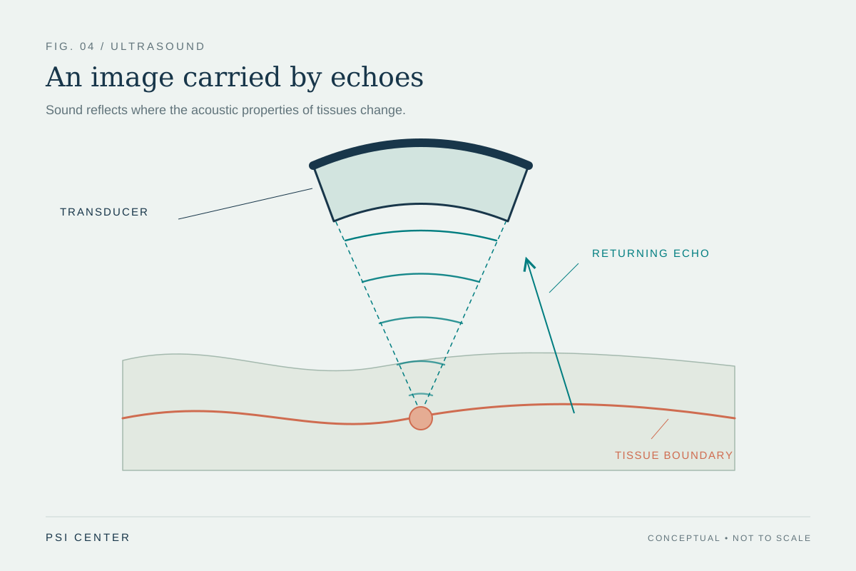
A probe array contains many transducer elements, and the scanner records a separate signal from each receive channel. For a chosen point in the body, it predicts when an echo should reach each element. Arrival times differ because the elements sit in different positions. Aligning the channel signals in time lets echoes from that point reinforce when added. This operation is called beamforming. [2]
Imagine a choir recorded from several seats: a sound arrives at each microphone at a slightly different moment. Shift the recordings to compensate, and that sound lines up; sounds from elsewhere usually do not. This is the basic timing idea, not a perfect filter. Tissue scatters sound, echoes overlap, and predicted travel times may be imperfect.
Delay-and-sum in a worked example
In delay-and-sum (DAS), the scanner selects a point, calculates a travel-time offset for each element, shifts the channel signals by those offsets, and adds them. It repeats this for many positions to form an image line or region. Pulse-echo imaging includes an outward trip and a return trip; echo time therefore encodes depth. The scanner uses an assumed sound speed to convert time into distance. [1] [2]
Suppose a target is about 4 cm beneath the probe and use 1,540 m/s as an approximate soft-tissue sound speed. The 8 cm round trip takes 0.08 m ÷ 1,540 m/s, or about 52 microseconds. This estimates total return time, not the small correction between neighboring elements. Those corrections come from geometry: an element nearer the target has a shorter path than one farther away. Compensating for those path differences helps the signals add coherently.
Focusing and steering use different delay patterns
To focus reception at a point, the scanner applies element-specific delays that compensate for the curved wavefront arriving from it. To steer to one side, it uses a progressive timing pattern across elements, making the combined response most sensitive to echoes from an off-axis direction. Transmit focusing shapes where emitted sound is strongest; receive focusing applies timing to echoes after they return. They are related but distinct operations. [2]
Electronic timing lets an array scan without mechanically moving one element through every line. The scanner repeats transmissions and receive calculations over locations or angles, then maps echo amplitudes into an image. Plane-wave methods use simplified transmission and can collect data quickly; combining multiple angles changes what data are acquired and how much processing is needed. [3]
The aperture is a finite listening window
The aperture is the set or width of elements used together. A wider active aperture can narrow the main response under some conditions, helping separate nearby reflectors. But an array is finite and its elements have spacing. Side lobes and grating lobes—secondary regions of sensitivity—can admit echoes from outside the intended beam. DAS cannot erase every such contribution. Element size and pitch constrain the patterns an array can form. [2] [3]
A beamformer is not a camera lens that focuses perfectly at every depth. Beam width varies with depth and frequency, while tissue affects the returning field. If the assumed sound speed differs from effective propagation along a path, predicted delays can be wrong and signals may not align. Real tissue is not a uniform block with one exact speed everywhere, so reconstruction remains a model-guided estimate from measured channel data.
Why sharper-looking images need careful scoring
Adaptive beamformers use the received data to change channel weights. Some can suppress incoherent or off-axis signal and make a target look narrower in a test image. Yet quality measures can move in different directions. A 2021 review reported phantom comparisons in which several adaptive approaches improved lateral resolution or contrast while contrast-to-noise ratio declined for some methods. This is method- and setup-specific, but “sharper” alone is not a complete performance claim. [2]
A fair comparison states what was measured and under which conditions. Point targets can test response width; contrast measures target-background difference; contrast-to-noise ratio includes variability. Phantoms isolate properties, while living tissue adds motion, heterogeneous sound speed, reverberation, and anatomy. The 2024 review also emphasizes quantitative assessment because adaptive processing can change image characteristics. An attractive example image is a reason to ask for validation, not proof of better clinical decisions. [3]
From channel signals to clinical meaning
The scanner displays processed data, often mapping echo magnitude to brightness. Beamforming determines how channel signals combine at a location, but later gain, compression, filtering, and display settings also affect appearance. A bright region does not simply mean “more tissue,” and a dark region is not automatically fluid or disease. Interpretation depends on imaging mode, anatomy, acquisition, and the question asked. [1] [2]
The useful model is a chain: the array emits sound, tissue returns echoes, each element records a signal, timing corrections align selected contributions, and processing turns the combined data into an image. Beamforming shapes which echoes add strongly and where they are assigned. It does not independently identify a condition, and an improved image metric does not establish improved care. Such claims need evidence beyond the physics of steering and focusing.
Explore the physics: an explanatory diagram
Sources & further reading
- Ultrasound National Institute of Biomedical Imaging and Bioengineering
- Advances in ultrasonography: image formation and quality assessment Journal of Medical Ultrasonics (2021)
- Beamforming methods in the field of medical ultrasonics IEICE ESS Fundamentals Review (2024)
Sources checked: 18 November 2026. The calculations illustrate physical models; they are not clinical settings.
Prepared for the independent PSI Center reading library. About edition dates and source checking.
This explainer describes scientific principles. It does not assess individual treatment choices.