How can echoes from a moving sound probe become a three-dimensional image? A conventional ultrasound scan estimates boundaries along a beam and displays a two-dimensional slice. To show a volume, the system needs spatial information from multiple locations, then a computer must organize those measurements into a coherent representation.
Echo time gives distance
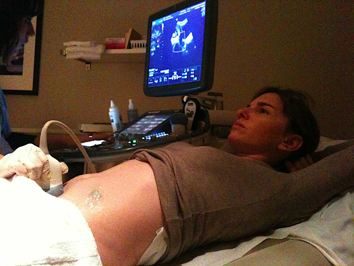
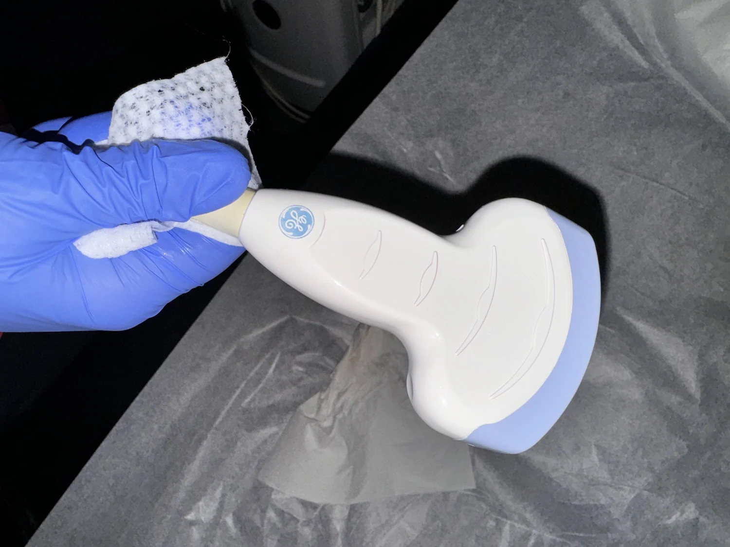
An ultrasound transducer sends sound waves into the body and detects echoes reflected from boundaries between tissues. The scanner uses the speed of sound and each echo's return time to estimate the boundary's distance from the probe [1]. A single beam gives information along a line; many neighboring beams can be assembled into a two-dimensional cross-section.
The echoes differ because tissue boundaries reflect sound differently. Gel placed between skin and probe helps prevent air pockets that could block sound from entering the body [1]. The transducer's active piezoelectric elements convert electrical energy into sound and returning sound into electrical signals [1]. These signals form the raw measurements from which the scanner creates an image.
A volume needs spatial sampling
To make a three-dimensional image, the system must know where measurements belong in more than one plane. One way to picture this is sweeping a thin sheet of light across a sculpture and recording each cross-section: the slices can be stacked if their positions are known. Ultrasound systems similarly combine spatially sampled information, whether the probe is moved or the transducer captures a volume, to represent depth and lateral position together.
The computer processes echo data and maps signal locations into a volume. A 3D display can then be viewed from different angles, much as a stack of slices can be digitally explored. The image is not a literal photograph of sound reflecting from every microscopic point. It is a reconstruction based on detected echoes, their timing, and the system's assumptions about sound travel.
Motion adds another dimension
Ultrasound can display images in 2D, 3D, or 4D, where 4D means 3D in motion [1]. Motion requires repeated volume information over time. For example, a moving fetus or a beating heart changes position while the system gathers and displays data. A sequence of volumes can convey movement, but the result depends on how quickly the data are acquired and how the system handles motion between measurements.
Functional ultrasound adds information such as movement and velocity of tissue or blood to anatomical images [1]. Doppler methods can display blood-flow speed and direction, while elastography maps relative tissue stiffness [1]. These measurements can be layered or interpreted alongside anatomical structure, but they answer different questions from simple shape. A colored area may represent a measured property rather than the tissue's natural color.
The reconstruction has limits
Sound does not travel equally well through every part of the body. Ultrasound is not good for imaging bone or air-filled tissues such as lungs, though there are special circumstances in which some bone or fluid-associated lung structures can be imaged [1]. An obstacle can block or redirect sound, leaving incomplete information behind it. A 3D rendering cannot recover details that the probe never measured clearly.
Computational models can simulate complex systems by combining mathematical and physical rules, and researchers use them to explore conditions that are difficult to test directly [2]. Ultrasound image reconstruction is a specific imaging task, while a research model may simulate biological behavior more broadly. In either case, the output depends on inputs and assumptions. Three-dimensional ultrasound is therefore best understood as a spatially organized interpretation of echoes, with its view shaped by the sound path, the sampling, and the reconstruction.
Explore the physics: an explanatory diagram

Sources & further reading
- Ultrasound NIBIB
- Computational Modeling NIBIB
Prepared for the independent PSI Center reading library. About edition dates and source checking.
This explainer describes scientific principles. It does not assess individual treatment choices.