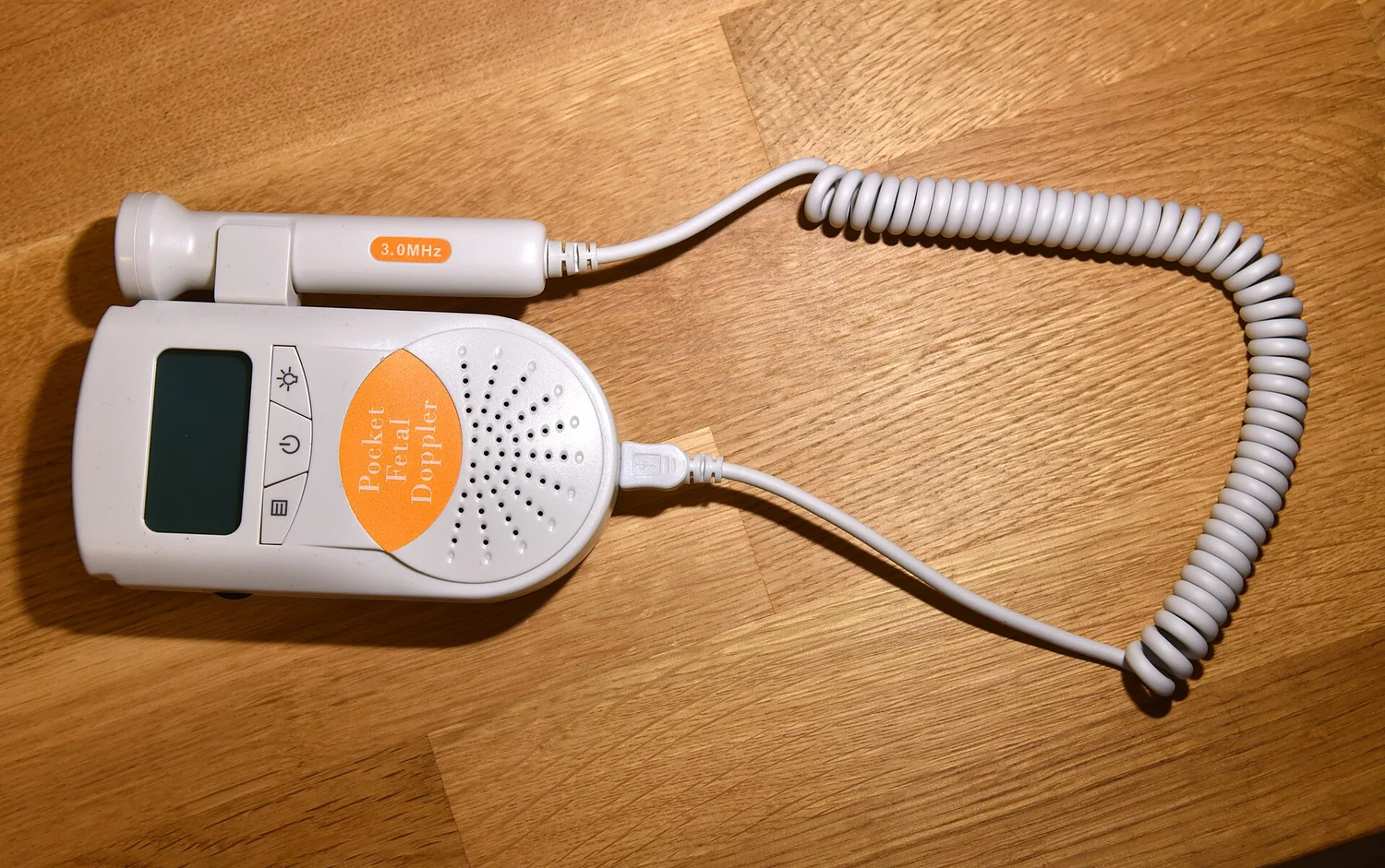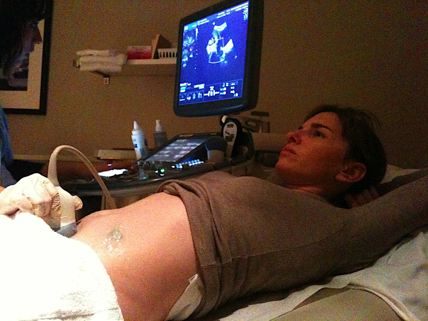
How can a probe resting on the chest show a heart valve opening and blood moving through a chamber? Echocardiography sends high-frequency sound into the body and listens for echoes returning from boundaries between tissues. A computer turns the timing and strength of those echoes into images. The method lets clinicians observe structure in motion, bringing the heart’s pumping action into view rather than presenting only a still outline.
A pulse travels, then echoes return
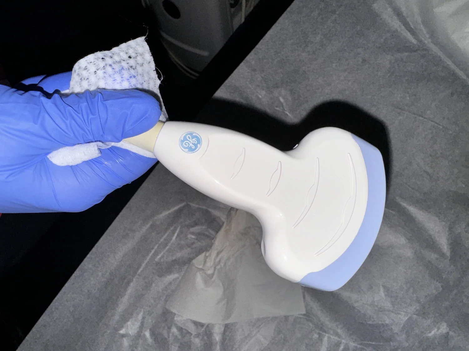
The sound used in diagnostic ultrasound is above the range of human hearing, typically at frequencies in the megahertz range. A probe called a transducer sends pulses into the body. When sound reaches a boundary between materials with different acoustic properties, some energy reflects back toward the probe [1].
The transducer detects each returning echo. Because sound travels at a known speed through tissue, the scanner uses the echo’s return time to estimate how far away the reflecting boundary is. Repeating this process at many depths creates a two-dimensional image of internal structures [1].
The probe converts between electricity and sound
Many ultrasound transducers use piezoelectric materials. These materials change shape slightly when an electric field is applied, producing a sound wave. In reverse, pressure from a returning echo creates an electrical signal in the material. The scanner sends pulses and then listens for these signals, switching the same probe between transmitting and receiving [1].
A layer of gel on the skin helps remove tiny air pockets between probe and skin. Air strongly interferes with sound transmission, so gel improves the acoustic connection. For heart imaging, the probe can be moved across the chest to collect views from different positions, each showing the heart from a distinct angle.
Watching chambers, valves, and motion
An echocardiogram can show heart chambers and valves as they move through the pumping cycle. The heart’s valves keep blood flowing in the right direction, while coordinated contractions move blood through the circulatory system [2]. Seeing the structures in motion lets an examiner relate anatomy to function, such as whether a valve opens and closes as expected.
The heart is not simply a hollow pump: it is layered tissue with an electrical system that controls the rate and rhythm of its beat [2]. An ultrasound image records echoes from moving structures over time. This makes it possible to observe motion directly, rather than infer every mechanical detail from a single still image.
Echoes can reveal blood flow too
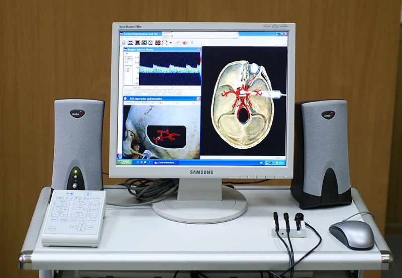
Functional ultrasound adds information about movement and velocity to anatomical images. Doppler methods use changes in the returning sound signal associated with moving blood cells to estimate flow speed and direction. Color Doppler displays this information as a map over the image, helping visualize flow in the heart and vessels [1].
Ultrasound has limits as well as strengths. It does not image well through bone or air-filled tissue, although it can be useful in selected situations where fluid provides an acoustic window [1]. Diagnostic ultrasound does not use ionizing x-rays, but its energy can have biological effects under some settings, so devices operate within accepted limits and use is guided by medical need [1].
Explore the physics: an explanatory diagram

Sources & further reading
- Ultrasound NIBIB
- How the Heart Works NHLBI
Prepared for the independent PSI Center reading library. About edition dates and source checking.
This explainer describes scientific principles. It does not assess individual treatment choices.