How can an ultrasound image show blood moving through a vessel? The probe sends sound into the body and listens for echoes. When sound reflects from moving blood cells, the returning frequency shifts slightly. By measuring that shift, a scanner can estimate whether blood is moving toward or away from the probe and how fast it is moving along the beam.
An echo carries motion information
Diagnostic ultrasound begins with a transducer, a device that sends out high-frequency sound and detects returning echoes. Sound reflects at boundaries between tissues, and the time taken for each echo to return helps the scanner estimate depth. The same probe can detect sound reflected from moving red blood cells. Their motion changes the frequency of the returning echo through the Doppler effect [1].
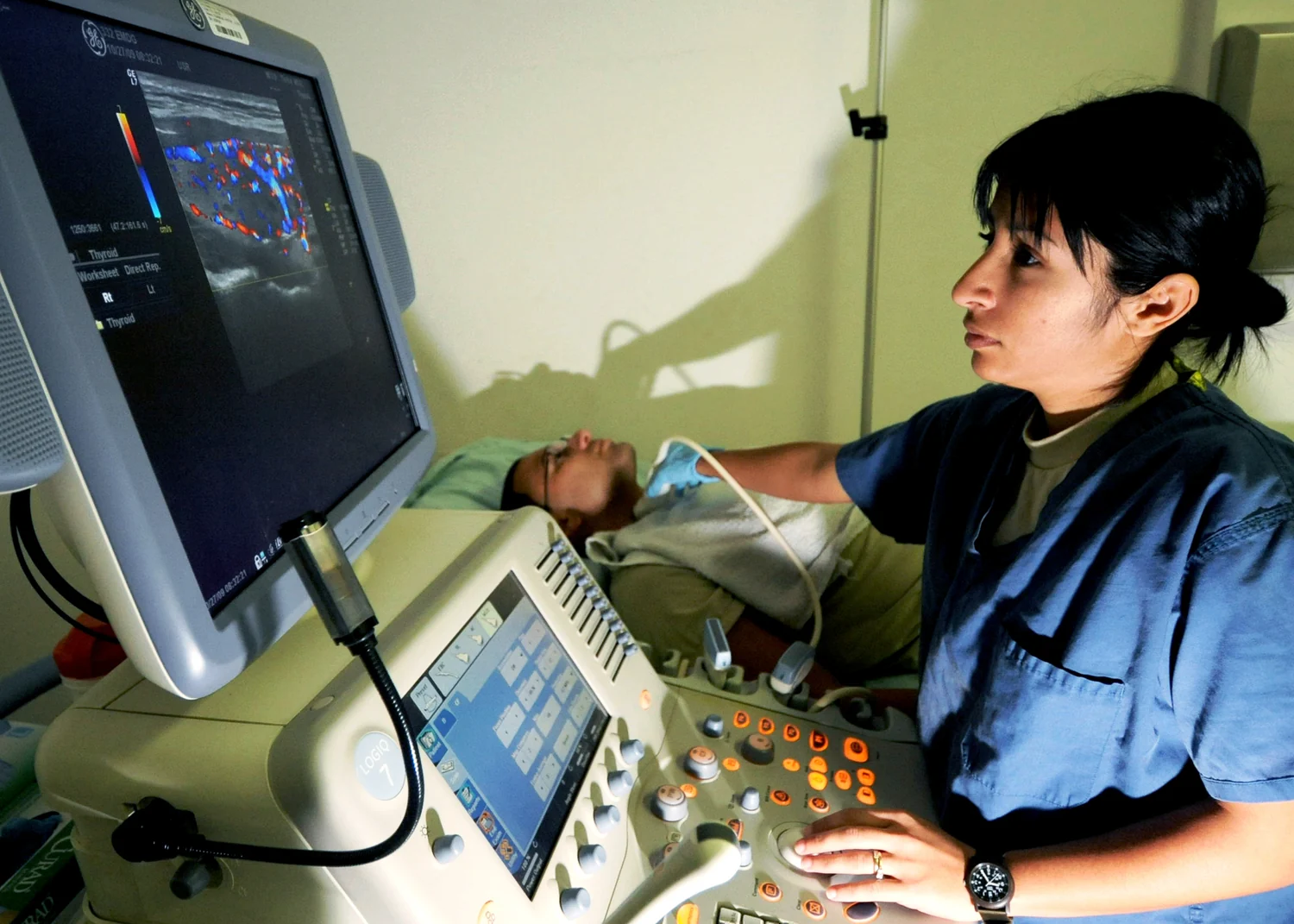
The Doppler effect is familiar from a passing siren: the sound seems higher as the source approaches and lower as it recedes. With ultrasound, the blood cells act as moving reflectors. The size and sign of the frequency shift depend on their velocity component along the sound beam. The scanner processes this shift to estimate flow direction and speed, then can display the result as a waveform or a color-coded map [1].
Angle is part of the measurement
Suppose blood flows directly toward the probe. Its motion is aligned with the beam, so the measured shift is relatively large. If the vessel lies almost perpendicular to the beam, much less of the motion is toward or away from the probe, and the frequency shift becomes small even if blood is moving quickly. The scanner is therefore measuring a component of velocity, not an angle-free speed in every circumstance.
This geometric dependence matters when interpreting Doppler images. The color map is a visual encoding of measured motion relative to the probe and chosen display settings; red and blue do not universally mean oxygen-rich and oxygen-poor blood. A waveform can show how the signal changes over a heartbeat, but it too depends on where and how the sample is taken. Functional ultrasound combines movement and velocity information with anatomical images to help visualize how a structure works [1].
From vessel map to clinical question
The heart pumps blood through a network of vessels that carries oxygen and nutrients to organs [2]. Doppler ultrasound can examine flow in vessels and the heart, helping clinicians assess direction and speed. One use is evaluating whether plaque buildup in the carotid arteries is obstructing blood flow toward the brain [1]. The measurement adds a dynamic layer to an image of vessel anatomy.
A useful mental experiment is to imagine measuring traffic from a roadside microphone. A loud, rapidly changing signal may indicate movement, but it does not by itself tell you every vehicle’s route or why traffic is slowing. Similarly, a Doppler finding must be interpreted with the vessel’s anatomy, the clinical question, and the broader exam. A map of motion is evidence about flow, not a complete diagnosis on its own.
Why ultrasound suits flow—and where it struggles
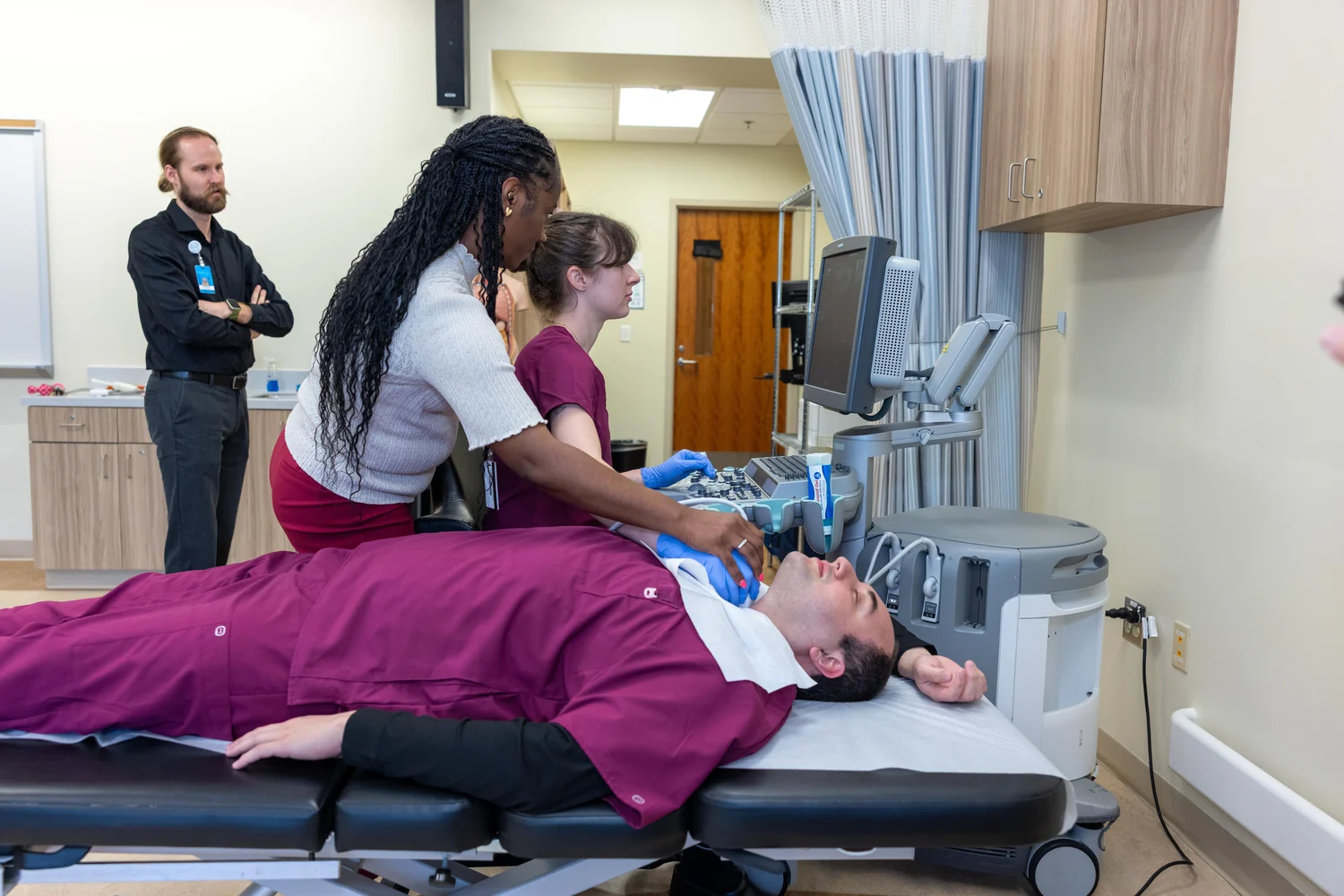
Ultrasound provides real-time echoes without ionizing x-rays, and Doppler extends that capability to moving blood. Gel between the probe and skin helps prevent air pockets that would block sound from entering the body [1]. The method can be used at the bedside and paired with anatomical imaging, making it a versatile way to inspect circulation in accessible regions.
Sound does not pass equally well through every material. Ultrasound is not good at imaging through bone or air-filled tissue, and measurements can be affected by probe position, beam angle, motion, and the quality of the acoustic window [1]. These limits explain why Doppler is one tool among several rather than a universal flow detector. Its distinctive contribution is to turn a returning sound wave into a velocity clue, linking the physics of echoes to the movement of blood.
Explore the physics: an explanatory diagram

Sources & further reading
- Ultrasound NIBIB
- How the Heart Works NHLBI
Prepared for the independent PSI Center reading library. About edition dates and source checking.
This explainer describes scientific principles. It does not assess individual treatment choices.