How does a scanner show a heart beating or a needle advancing inside the body as it happens? It listens for echoes. A probe sends high-frequency sound into tissue, then measures returning reflections and their timing. Because this exchange happens repeatedly, the image can update as anatomy moves. The result is not a photograph of a frozen interior, but a sequence of measurements that can reveal motion while also helping guide a procedure.
Reading a returning echo
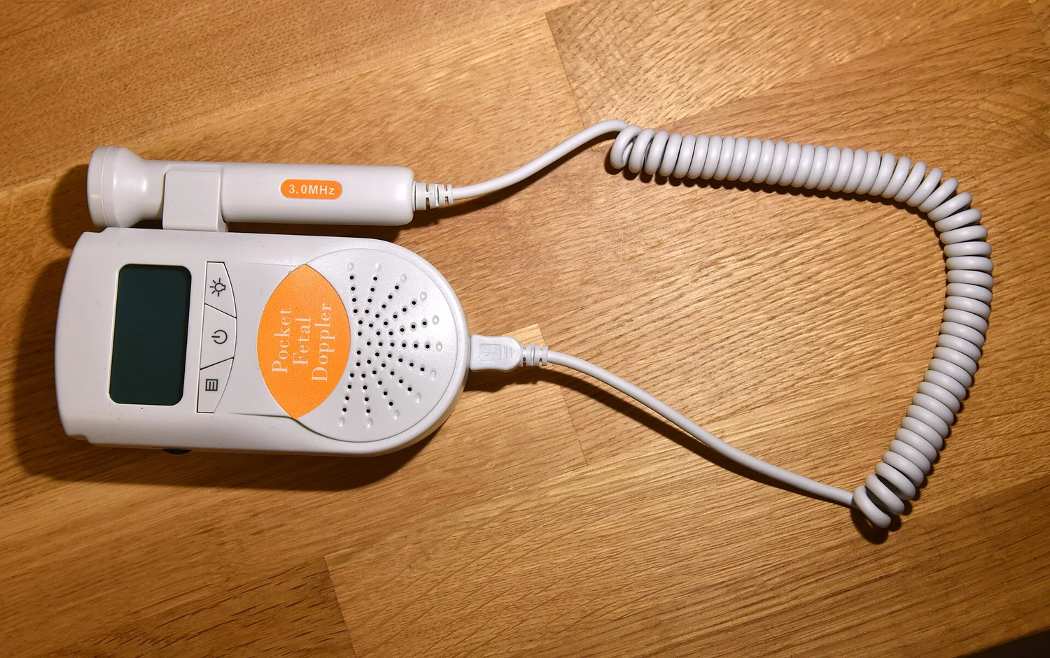
A transducer, or ultrasound probe, both emits sound waves and detects echoes. Many probes use piezoelectric materials: applying an electric field makes them vibrate and produce sound, while returning sound can generate an electrical signal. The scanner estimates how far away a tissue boundary lies from the time an echo takes to return and the known speed of sound in tissue. Echo strength adds information about the reflecting structure [1][2].
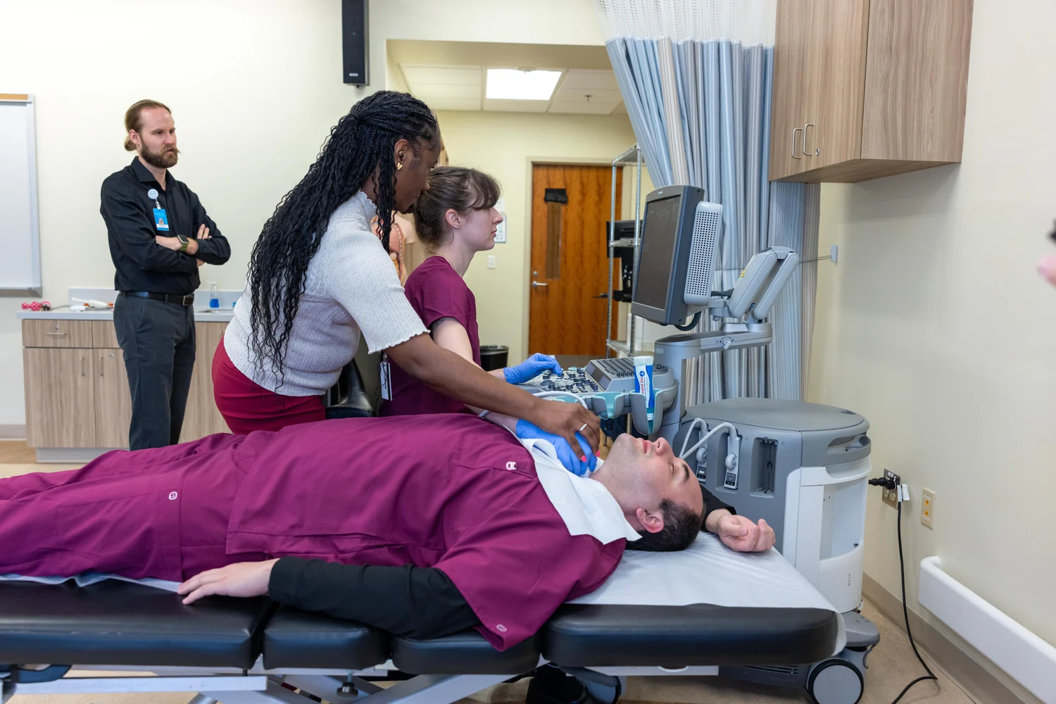
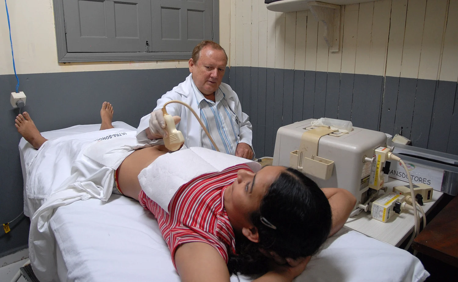
A thin layer of gel helps couple the probe to skin. Air pockets between the probe and skin would interrupt the sound path, because ultrasound does not pass readily through air. Inside the body, reflections arise at boundaries where tissue properties change, such as between fluid and soft tissue. The machine repeatedly sends pulses and processes echoes, turning those distances and strengths into images that can be refreshed as the body changes [1].
Motion becomes part of the image
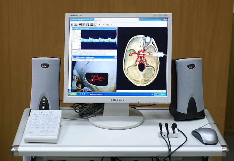
A still image can show where an organ is; a real-time sequence can show how it moves. Ultrasound imaging can display internal organs in motion and blood flowing through vessels. Doppler methods use shifts in the returning sound to estimate blood-flow direction and speed, while color Doppler presents flow information as a map over anatomy [2][1].
Consider a clinician guiding a needle toward a visible target. The needle and surrounding anatomy can shift as the instrument advances or tissue deforms. A live ultrasound view lets the operator see the needle’s position relative to the target during the procedure, rather than relying only on a scan made earlier. Ultrasound is used to guide biopsies and needle placement, and to view a catheter tip as it travels within a blood vessel [1][2].
Useful windows, blind spots
Sound does not form equally useful images through every material. Ultrasound is generally poor at imaging bone and tissues containing air, including the lungs. Bone can block or strongly reflect the waves; air disrupts the path. Under some conditions, such as when fluid is present, parts of the lung lining can be imaged, but ultrasound is not a universal way to look through the body [1].
Soft tissue and fluid often offer more workable paths, which helps explain why ultrasound is used for abdominal organs, the heart, blood vessels, muscles, and pregnancy imaging. The probe’s position and angle also matter: changing either can change which boundaries return strong echoes. A live image is thus a view from a particular acoustic window, not a complete map of every structure in the body.
Imaging sound and treatment sound
Diagnostic ultrasound is designed to gather information; therapeutic ultrasound is designed to affect tissue. High-intensity focused ultrasound, or HIFU, concentrates acoustic energy so that tissue can be heated or destroyed. Some HIFU uses have FDA approval for specified purposes, while other possible applications remain under investigation [1]. A treatment beam’s purpose and intensity distinguish it from the echoes used to make a diagnostic image.
Ultrasound does not use ionizing radiation like X-rays, but that does not mean sound energy has no physical effects. It can heat tissue slightly and, in some conditions, produce small gas pockets called cavitation. Prudent medical use aims to keep exposure as low as reasonably achievable while preserving diagnostic quality; unnecessary prolonged exposure and use by untrained operators raise particular concerns [2].
Explore the physics: an explanatory diagram

Sources & further reading
- Ultrasound NIBIB
- Ultrasound Imaging FDA
Prepared for the independent PSI Center reading library. About edition dates and source checking.
This explainer describes scientific principles. It does not assess individual treatment choices.