Can a flash of light make sound inside the body? In photoacoustic imaging, it can. A brief laser pulse is absorbed by tissue, producing a tiny rapid temperature change; the resulting expansion launches an ultrasound wave that a detector can record. The method uses light to start the signal and sound to carry it back out.
Absorption turns light into heat
Light entering tissue may be absorbed, scattered, reflected, or transmitted. In photoacoustic imaging, the useful event is absorption: molecules such as those in blood take up some of the optical energy. Because different tissues and molecules absorb light differently, the pattern of absorption can provide contrast about what is present and where. [1]
The laser pulse is brief, so absorbed energy arrives faster than the tissue can readily dissipate it. The small temperature rise makes the absorbing region expand slightly. That quick expansion pushes on neighboring tissue and generates a pressure wave. The wave is ultrasound: sound at frequencies above ordinary human hearing, traveling through tissue as mechanical motion.
Expansion makes an acoustic pulse
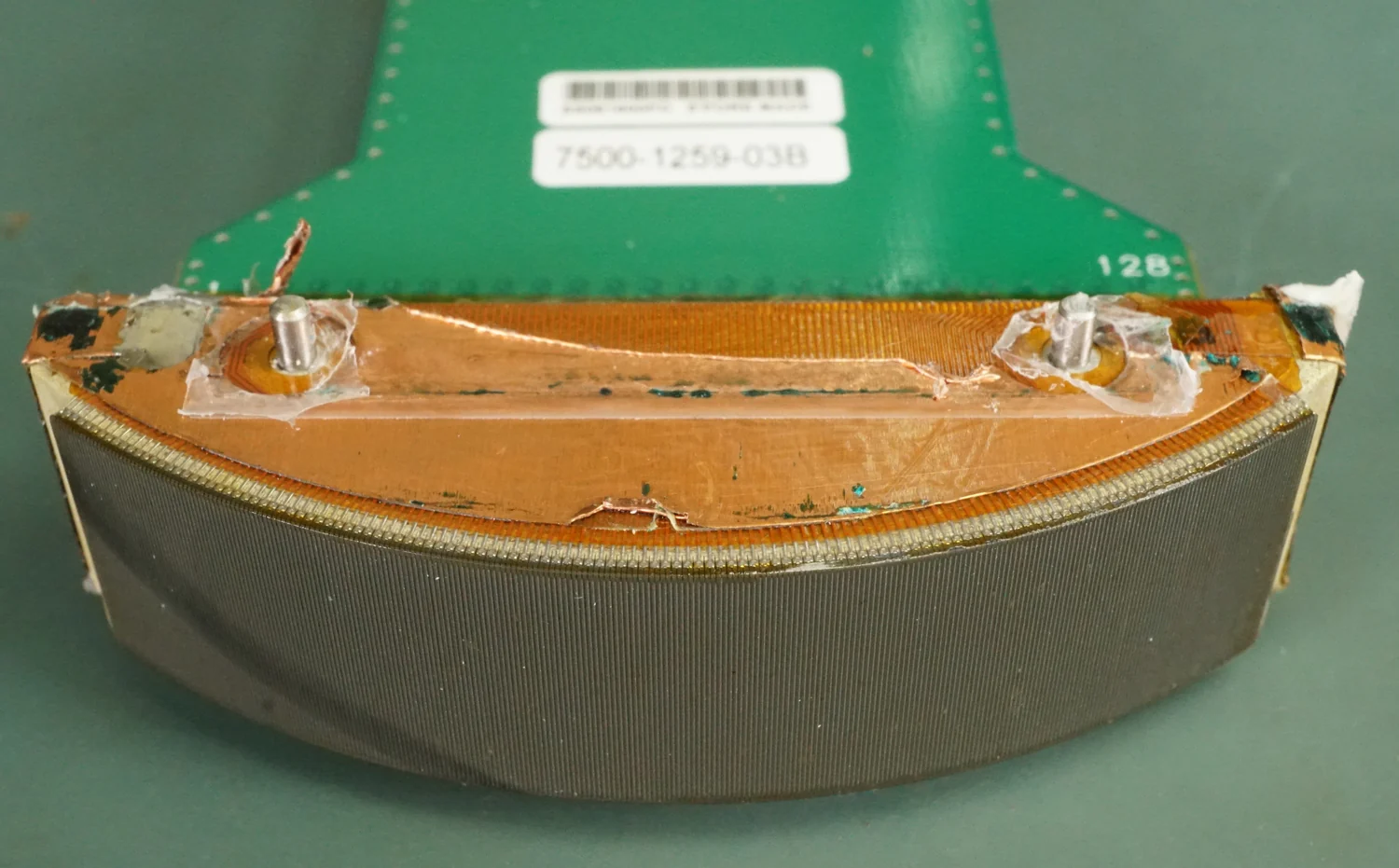
Think of a tiny region of tissue as a springy patch. A short pulse warms it; it expands; then it settles back. That rapid change launches pressure disturbances that move outward. A photoacoustic transducer detects the returning waves and converts them into electrical signals. From their timing and shape, reconstruction methods estimate where the original absorption occurred. [1]
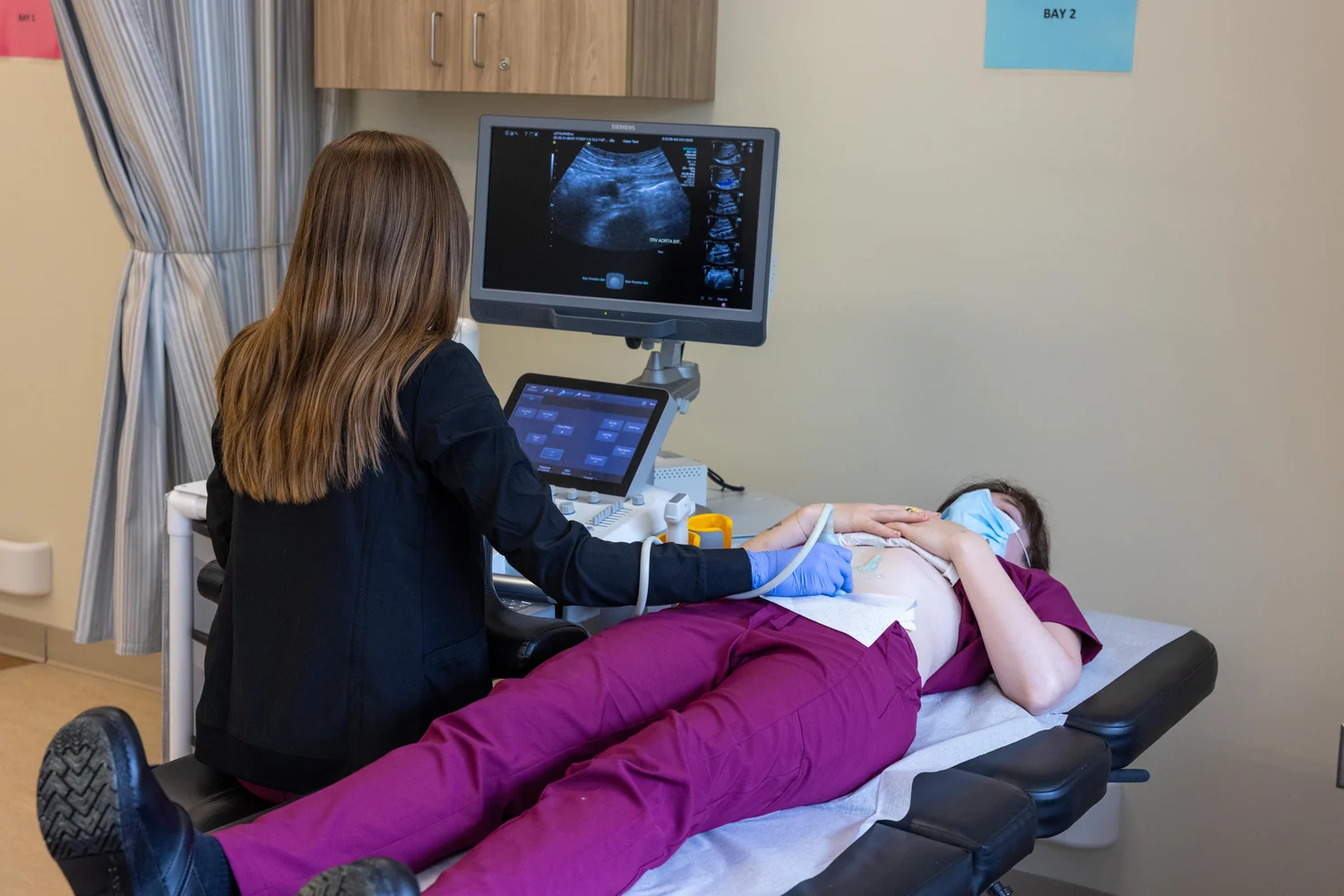
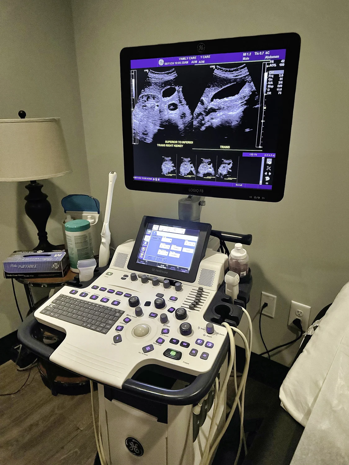
This is different from ordinary diagnostic ultrasound, where a probe first sends sound into the body and listens for echoes from boundaries between tissues. In photoacoustics, optical absorption generates the sound inside the tissue, and the detector listens for that sound. The ultrasound part is still essential: it transports information from within tissue to sensors at the surface. [2]
Why combine optical contrast and sound?
Light can be sensitive to tissue chemistry and blood-related properties, but scattering makes it difficult to form sharp conventional optical images deep inside the body. Ultrasound generally travels farther through soft tissue and can be detected after it has emerged. Photoacoustic imaging therefore joins optical contrast with acoustic reach, creating a hybrid rather than purely optical technique. [1]
The result can reveal features such as blood vessels and oxygenation-related patterns. NIBIB describes photoacoustic imaging applications that include monitoring vessel growth in tumors, detecting skin melanomas, and tracking blood oxygenation in tissues. These are imaging uses and areas of research; a displayed feature still needs interpretation in context and does not alone establish a disease diagnosis. [1]
A signal is not a complete picture
The process depends on both the light reaching absorbers and the ultrasound reaching the detector. Optical scattering, tissue depth, acoustic attenuation, and the detector geometry all shape the signal. A reconstruction is an estimate based on measured waves, not a direct snapshot of every molecule. The modality’s strength is the physical chain that converts absorption into sound; that chain also defines what can be measured.
Photoacoustic systems are being developed for biomedical research and potential clinical applications. One NIBIB-supported approach has imaged blood vessels and estimated flow speeds in human hands and feet, while work on broader applications remains an active research area. [1] The basic idea remains strikingly simple: light deposits energy selectively, rapid expansion creates pressure, and ultrasound brings that pressure pattern back to a sensor.
Explore the physics: an explanatory diagram

Sources & further reading
- Optical Imaging NIBIB
- Ultrasound NIBIB
Prepared for the independent PSI Center reading library. About edition dates and source checking.
This explainer describes scientific principles. It does not assess individual treatment choices.