Why can an ultrasound probe show fine detail near the skin yet struggle to see structures deeper inside? The answer begins with sound’s wavelength. Raising its frequency shortens that wavelength, helping distinguish small features, but higher-frequency sound is also absorbed and scattered more readily in tissue. A scan therefore involves a practical trade: choose the detail needed at the target depth, then interpret the echoes that make the image.
Shorter waves resolve smaller details
Frequency is the number of sound-wave cycles passing a point each second. Diagnostic probes generally operate in the megahertz range, far above human hearing. At a given speed of sound, increasing frequency shortens wavelength: the wave’s repeating pattern occupies less distance. That makes it easier for an imaging system to distinguish nearby boundaries and small features, much as fine pencil strokes can outline a small drawing more precisely than broad strokes.
Imagine trying to map two tiny reflectors positioned close together. A longer wave may blur their echoes into one broad return; a shorter wave can produce more distinct echoes. This is a useful rule of thumb, not a guarantee that every small structure will appear. Image quality also depends on the scanner, the tissue, the angle of the beam and the strength of the returning signal. Ultrasound images are built from echoes at boundaries between tissues, whose acoustic properties differ [1].
Tissue gradually dims the beam
Sound does not travel through the body without loss. Some energy is absorbed and converted to heat; some is scattered away from the beam or reflected at boundaries. In general, attenuation—the reduction in wave strength during travel—increases with frequency in soft tissue. The returning echo must make the trip back to the probe too, so losses along the outward and return paths both matter.
Picture a flashlight beam crossing a hazy room: a brighter, finer beam may reveal detail nearby, while distance and haze weaken what reaches the far wall and what reflects back. Ultrasound is not light, but the comparison illustrates why a very high frequency is not automatically the best choice for a deep target. A weak echo can be difficult to distinguish from background noise, even if the short wavelength could in principle resolve a fine boundary.
Depth comes from echo timing
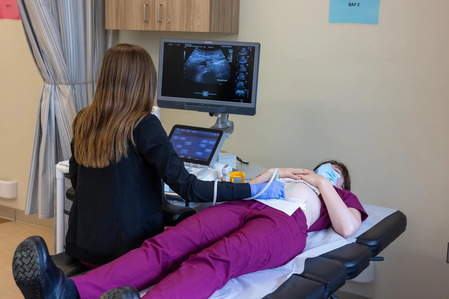
A transducer sends a brief pulse and then listens. Many probes use piezoelectric materials, which convert electrical signals into sound and returning sound into electrical signals. The scanner estimates how far away a reflecting boundary lies from the time taken for the echo to return, using an assumed speed of sound through tissue [1]. It assembles many such measurements into an image; brightness also reflects echo strength and the way the system processes signals [2].
This timing explains why an image is not a direct photograph. The machine infers position from returning waves, and echoes may overlap or weaken as they travel. Air pockets can block transmission, which is one reason gel is applied between probe and skin. Bone and air-filled regions also pose challenges for many examinations [1]. Frequency changes how much detail can be resolved and how far usable echoes tend to travel, while timing converts those echoes into depth.
A choice shaped by the target
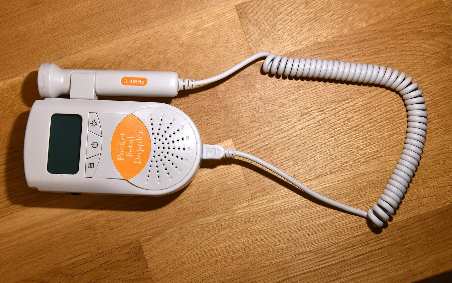
For a structure close to the skin, fine detail may matter most, so a higher-frequency probe can be useful. For a deeper organ, stronger penetration may matter more, making a lower frequency a sensible trade. The operator chooses a probe and settings for the anatomy and question at hand; one frequency cannot maximize both fine detail and depth in every situation.
Diagnostic ultrasound does not use ionizing X-rays, but it sends energy into tissue. Ultrasound can produce slight heating and, under some conditions, mechanical effects such as small gas pockets; regulators recommend prudent use while maintaining diagnostic quality [2]. This is one reason scanning is a clinical imaging technique rather than simply turning up power until every feature appears. Frequency is a design choice within a broader process: transmit sound, collect echoes, and judge whether the resulting information is adequate for the target.
Explore the physics: an explanatory diagram

Sources & further reading
- Ultrasound NIBIB
- Ultrasound Imaging FDA
Prepared for the independent PSI Center reading library. About edition dates and source checking.
This explainer describes scientific principles. It does not assess individual treatment choices.