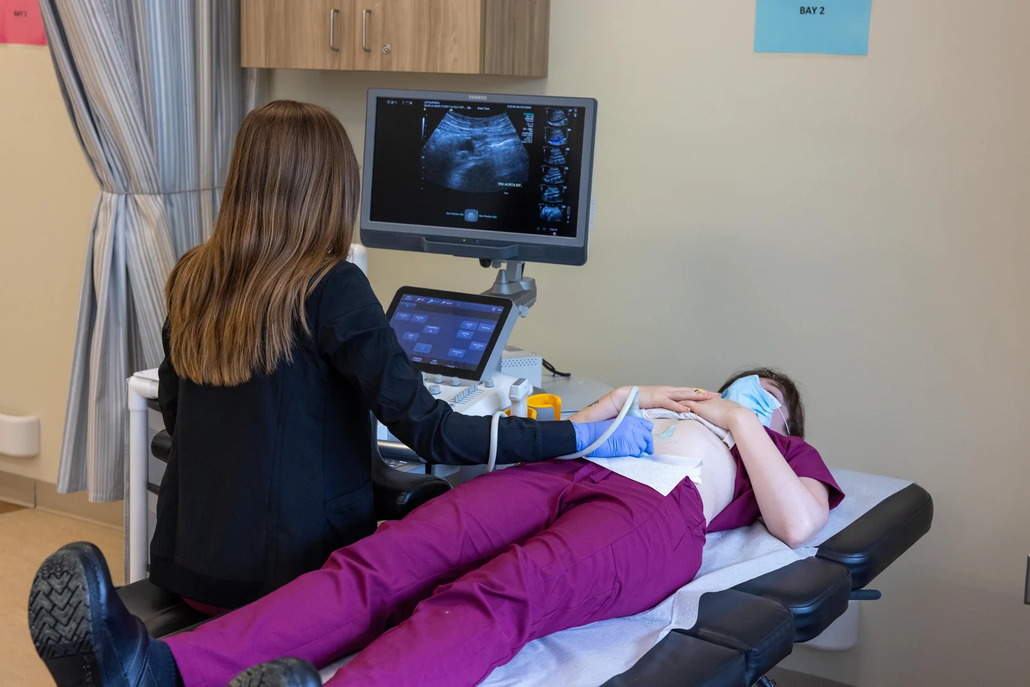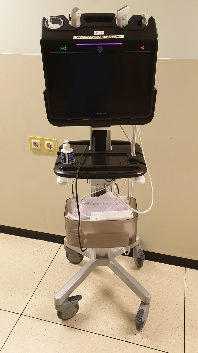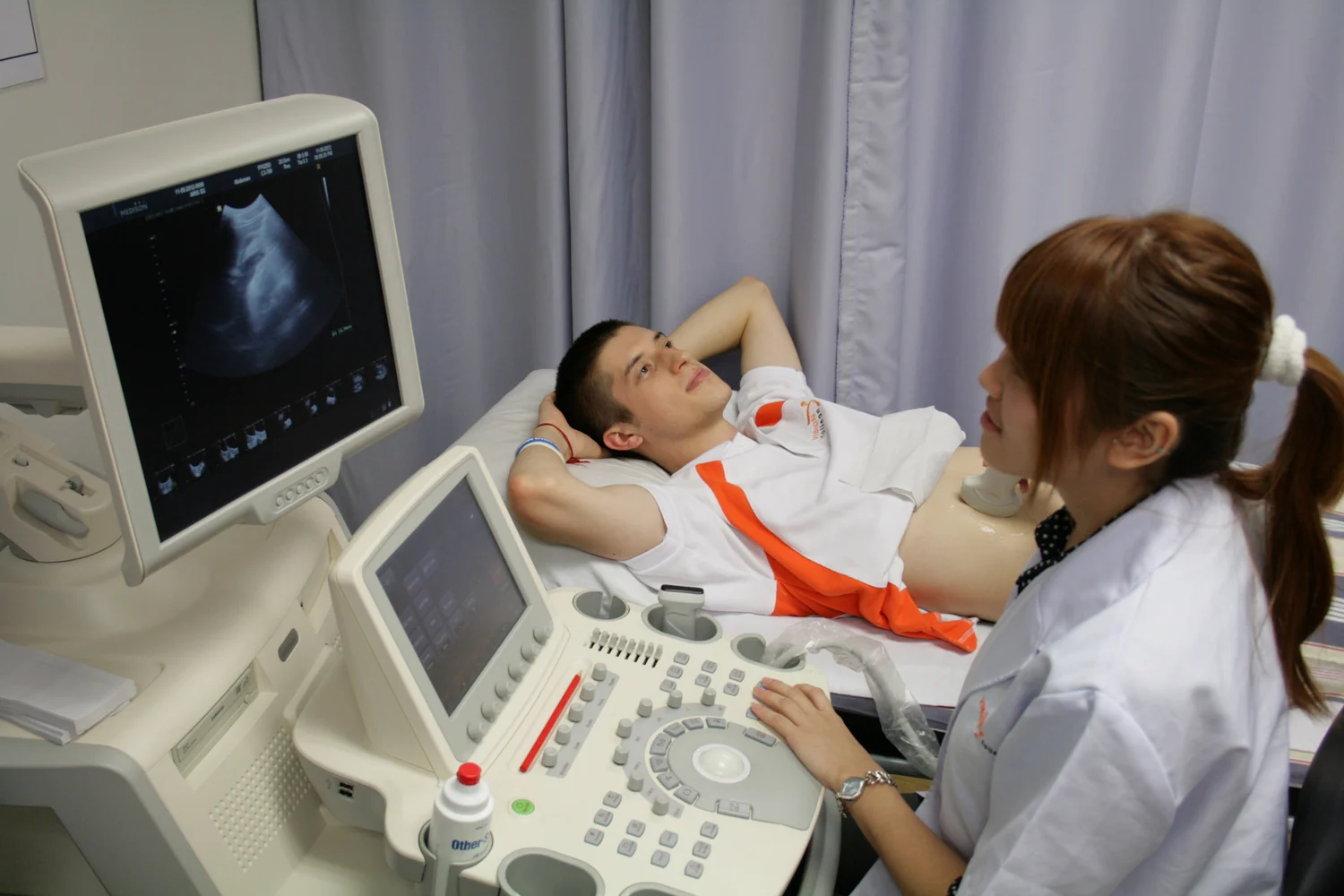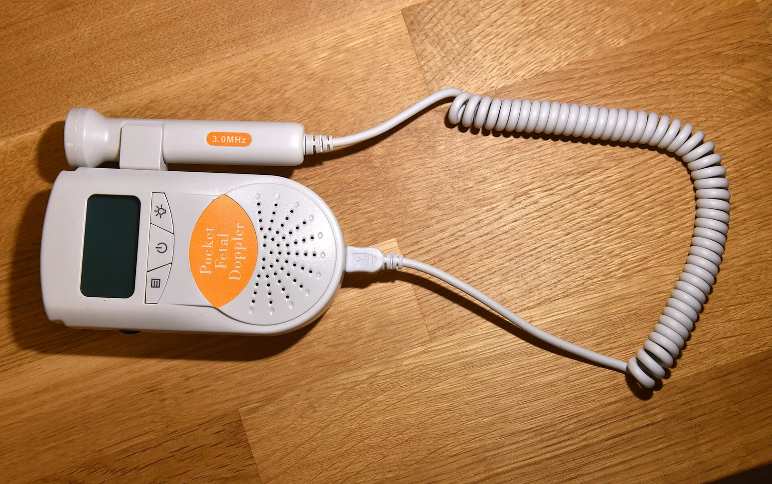
How can a handheld probe make a picture of something hidden beneath the skin? It sends sound into the body and listens for echoes returning from boundaries between tissues. The crucial component does both jobs: it creates a pulse, then detects the returning vibration. A scanner uses the timing and strength of those echoes to estimate where structures lie. The image is therefore a reconstruction from sound’s trip through tissue, not a photograph taken from inside the body.
A crystal with a two-way conversion
Many ultrasound probes contain piezoelectric ceramic elements. Piezoelectricity means that certain materials change shape slightly when an electric field is applied, and produce an electrical signal when pressure deforms them. In a probe, a rapid electrical pulse makes the element vibrate and launch a sound wave; an echo that returns later vibrates it in turn, creating a voltage the scanner can measure [1].
The same element can alternate between sending and receiving. The sound has a frequency above human hearing, typically in the megahertz range for diagnostic probes. The probe is not simply a tiny speaker paired with a separate microphone: its active material converts energy in both directions, which lets a compact device transmit a pulse and detect its reflections [1] [2].
A boundary sends part of the sound back
Imagine a sound pulse meeting the boundary between fluid and soft tissue. Some energy continues onward, while some reflects toward the probe. Similar echoes arise at boundaries between other tissues. How much reflects depends on the change in acoustic properties across that boundary; the returning signal is not a label saying “this is organ X,” but physical evidence that the wave encountered a change [1].
Contact matters because air can interrupt the sound path. A thin layer of gel fills small gaps between skin and probe, helping waves enter the body instead of reflecting at pockets of air. During an exam, the probe’s position and angle change which structures lie in the path and which echoes return. The FDA describes the image as based on reflections, signal amplitude, and travel time [2].
Turning travel time into depth
Sound travels through tissue at a speed the scanner can use as an estimate. If an echo takes longer to return, the reflecting boundary is generally farther from the probe. Because the wave makes a round trip, the scanner relates the elapsed time to an outward and return path, then estimates depth. Repeating this process for many pulses and directions builds a two-dimensional image of tissue boundaries [1].
Echo strength supplies another clue: a strong reflection may appear differently from a weak one. The scanner maps these signal differences into image brightness. This is an inference from measured sound, not a direct view of tissue. Structures that reflect similarly can be difficult to distinguish by a simple echo image, while motion or a changing probe angle affects what is captured [2].
The same sound can answer other questions
A basic anatomical scan maps structure, but ultrasound can also be used to examine motion. Doppler methods use changes in reflected sound associated with moving blood to estimate its direction and speed; color-coded maps can display flow. Elastography uses information about how tissue deforms or moves to estimate relative stiffness. These are different measurements layered onto the acoustic signal, not separate forms of radiation [1].
Diagnostic imaging and therapeutic ultrasound also have distinct aims. Imaging uses echoes to form pictures; therapeutic systems deliver higher acoustic output to heat, move, or destroy selected tissue. Diagnostic ultrasound does not use ionizing x-rays, yet sound energy can produce biological effects such as heating and, under some conditions, cavitation, or small gas pockets. FDA guidance therefore describes prudent, medically appropriate use rather than treating sound as energy-free [2].
Explore the physics: an explanatory diagram

Sources & further reading
- Ultrasound NIBIB
- Ultrasound Imaging FDA
Prepared for the independent PSI Center reading library. About edition dates and source checking.
This explainer describes scientific principles. It does not assess individual treatment choices.