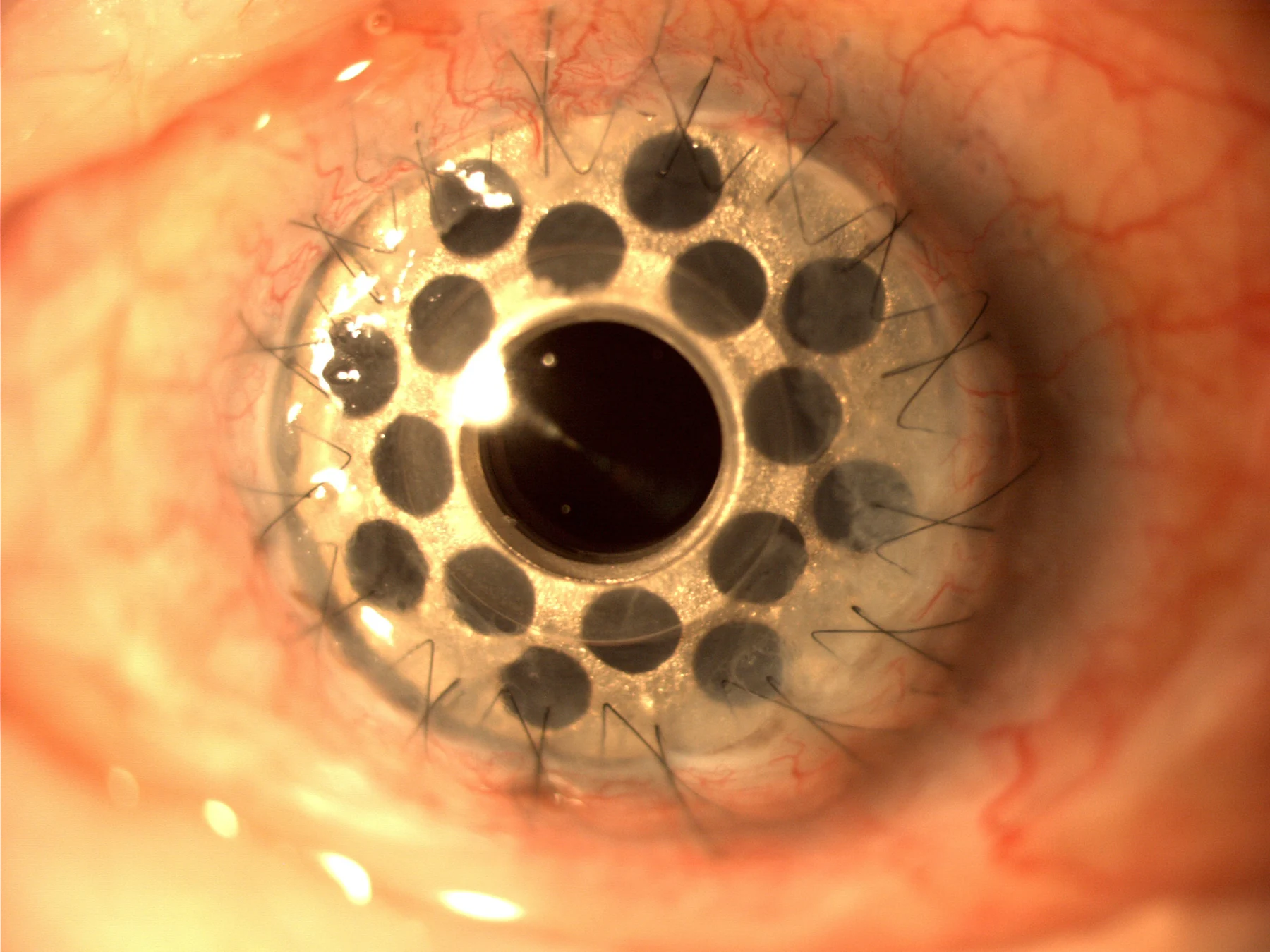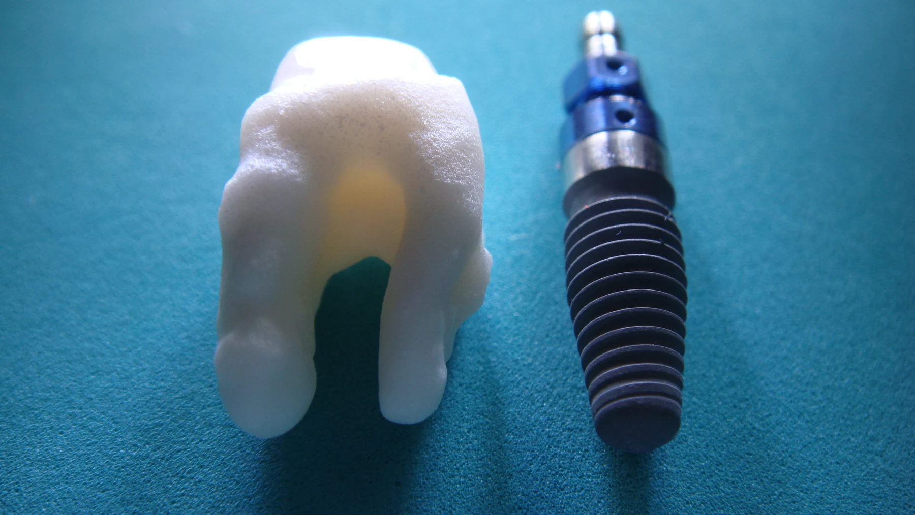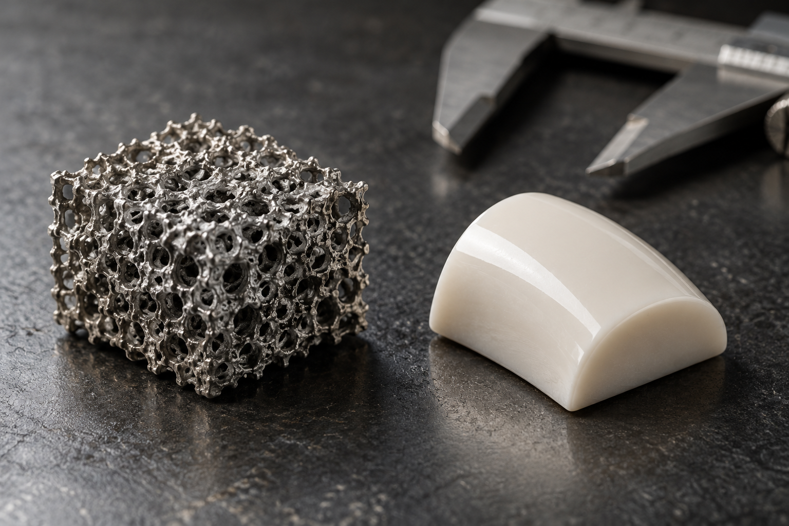
Why can two materials with the same ingredients behave differently after they are placed in contact with tissue? Their internal architecture may differ. A scaffold full of pores is not simply a solid with empty spots: pore size and the pathways linking pores govern how fluid moves, what can enter, and where cells encounter the material. Those geometric details influence function, though they do not by themselves prove a clinical benefit.
A material has an inner landscape
Biomaterials are engineered or adapted materials that interact with the body. They can take the form of foams, fibers, films, coatings, or structural frameworks intended to support tissue engineering [1]. A porous scaffold adds a network of open spaces within that framework. Its overall shape may be visible to the eye, while the pores and connecting passages are much smaller.
A useful mental picture is a sponge. Two sponges can have similar outer dimensions but different internal spaces: one may have broad cavities and narrow necks, another smaller cavities connected by wider channels. Water will move through them differently. In a biomaterial, the corresponding questions concern not just how much empty volume exists, but whether pores connect and how easily fluid can travel from one region to another.
Size changes access
Pore size affects which things can enter a space and how much room they have to move. A very narrow passage can restrict transport, while a larger opening may allow fluid and larger structures to pass more freely. In tissue, cells and molecules meet a material through this geometry. The scaffold’s architecture therefore helps set the physical conditions for contact, exchange, and tissue ingrowth.
Pore dimensions also interact with material properties such as stiffness and chemistry. Biomaterials may be designed to resemble aspects of the extracellular matrix, the natural molecular scaffolding around cells, in structure, stiffness, or composition. Matching some of these features can encourage integration or reduce aspects of the foreign-body response, though a design goal is not proof that every material will achieve it in patients [1].
Connectivity shapes transport
Connectivity asks whether neighboring pores form continuous routes or end in isolated pockets. Imagine pouring water onto a maze: large rooms are not useful for through-flow if narrow walls block the exits. In a scaffold, connected pathways can affect how fluids and dissolved substances reach deeper regions. Pores that do not link to the outside may contribute to measured void space but provide little route for transport across the material.
Computational models can help researchers study such complex systems by changing variables in a virtual environment and predicting how outcomes may shift [2]. A model might represent a scaffold as a network of pores and test how fluid movement changes when a passage narrows or a connection disappears. Such simulations can help identify useful experiments, but their predictions depend on assumptions and require comparison with laboratory or clinical evidence. The virtual material is a tool for asking focused questions, not a substitute for observing the real one.
Architecture is one part of performance
The right pore structure depends on the job. A material supporting load-bearing tissue must also withstand forces and repeated motion; a framework for soft tissue has different mechanical demands. Materials can be selected for strength, flexibility, wear resistance, degradation, or drug delivery, depending on their intended use [1]. Pore size and connectivity operate alongside these properties rather than replacing them.
Researchers study biological responses to materials because the body may degrade a foreign object or surround it with scar tissue, potentially affecting device function. Material structure can help shape those interactions, and tissue maps or models can reveal details that are hard to infer from an outer surface alone [1] [2]. Yet a promising architecture, an animal study, or a computational result does not establish that a biomaterial is clinically proven. The central engineering question is how a specific pore network, material, and application work together, followed by evidence that the resulting device performs safely and usefully in its intended setting.
Sources & further reading
- Biomaterial Technologies NIBIB
- Computational Modeling NIBIB
Prepared for the independent PSI Center reading library. About edition dates and source checking.
This explainer describes scientific principles. It does not assess individual treatment choices.