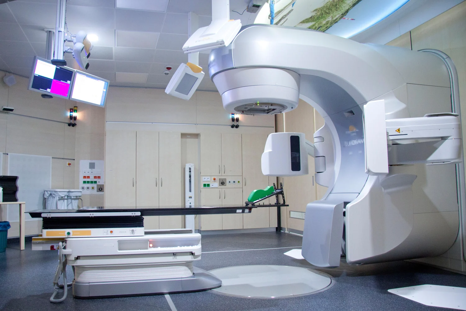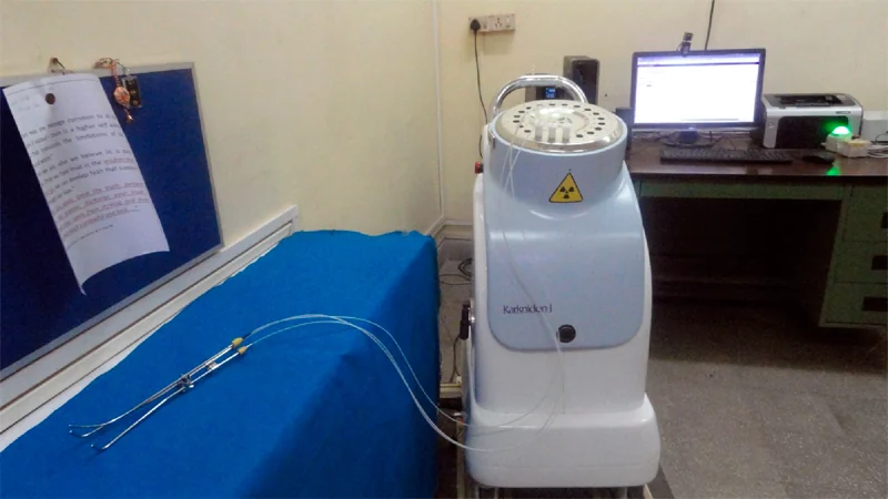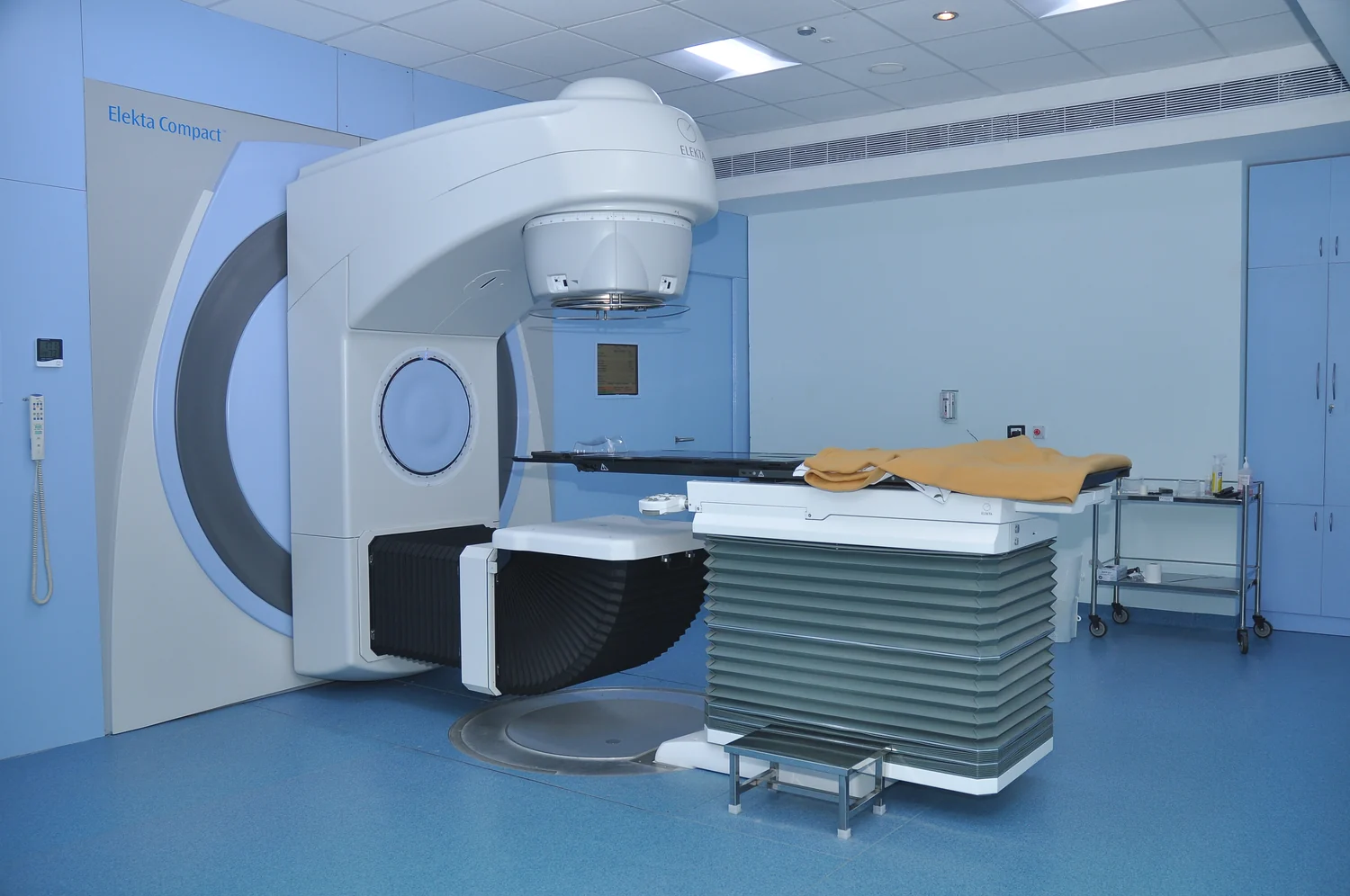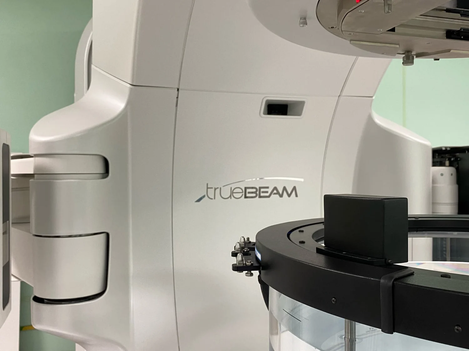
How can radiation reach a small tumor while limiting exposure to the tissue along the way? Stereotactic treatment approaches the problem geometrically: many beams enter from different directions, each contributing a small amount along its path and a concentrated dose where they converge. The method depends on more than a powerful machine. Images, positioning, and the target’s motion all shape how precisely that meeting point matches the intended anatomy.
A dose built from many paths
External beam radiation is a local treatment: a machine directs radiation at a specific region rather than treating the whole body. Radiation damages the DNA of cancer cells, disrupting their ability to divide or causing them to die. That process takes time; cells can continue dying for weeks or months after treatment ends. Nearby healthy cells can also be affected, which is why planning aims to treat the tumor while limiting dose to surrounding tissue [2].
Imagine shining several dim flashlights toward one spot on a wall. Each beam illuminates part of its route, but the overlap is brightest where they meet. Stereotactic radiosurgery uses the same geometric principle with focused, high-energy radiation beams: each small beam has little effect on the tissue it crosses, while their combined dose is concentrated at the target. The technique is used for small tumors with well-defined edges in the brain and central nervous system [1].
The target has to stay put
Precision begins before treatment. Planning images, including CT, MRI, or PET, help a computer map the tumor and calculate beam paths. A head frame or another positioning device helps keep a person still during stereotactic radiosurgery. Reproducing the planned position matters because even a small shift can move the convergence point relative to a small target [1].
For a thought experiment, picture trying to aim several narrow pointers at a dot on a sheet of paper. If the sheet moves after you set the pointers, they still converge, but no longer on the dot. Immobilization holds the body in the planned relationship to the machine. In image-guided radiation approaches, scans can also be taken during sessions to check for changes in tumor size or location and allow position or dose adjustments when needed [1].
Brain and body are different targets
Stereotactic radiosurgery usually treats small, sharply defined targets in the brain or central nervous system. Stereotactic body radiation therapy applies a similar idea to small, isolated tumors elsewhere, often in organs such as the lung or liver. Those organs move with ordinary actions such as breathing or digestion, so the target may not remain in the same place as it would inside a stabilized skull. Body treatments are therefore usually divided into more than one dose, sometimes up to five daily treatments [1].
That distinction is physical, not just a naming convention. A converging beam plan works best when the target remains where the plan expects it. With a moving target, treatment teams have to account for position changes, and the dose cannot be aimed as precisely as in stereotactic radiosurgery. The number and schedule of treatments vary with the cancer and the target’s location; the label alone does not determine a course [1].
Precision is a planning goal, not a force field
Radiation planning seeks to deliver the highest prescribed dose to the tumor while sparing normal tissue nearby. It cannot make the surrounding body disappear from the beams’ paths. With photon beams, some radiation scatters along the path and continues beyond the tumor. Proton beams behave differently: they can stop at the tumor, potentially reducing exposure to normal tissue, but comparisons with photon treatment remain a subject of clinical trials [1].
The word “stereotactic” therefore describes a way of localizing and delivering radiation, not a guarantee that healthy tissue receives none or that a particular treatment is suitable for every tumor. Tumor size, shape, location, nearby sensitive structures, and movement all matter. The underlying aim is carefully planned dose placement, while the biological effect—DNA damage and its consequences—unfolds over time [2].
Sources & further reading
- External Beam Radiation Therapy National Cancer Institute
- Radiation Therapy to Treat Cancer National Cancer Institute
Prepared for the independent PSI Center reading library. About edition dates and source checking.
This explainer describes scientific principles. It does not assess individual treatment choices.