How can a radiation beam reach a tumor without treating every healthy cell along the way? It cannot avoid all nearby tissue, so planning is a problem of geometry, dose, and biology. Clinicians use images to locate the target, choose how radiation enters the body, and shape the dose around it. The aim is to deliver an effective prescribed dose to the tumor while reducing unnecessary exposure to normal structures, not to make those structures invisible to radiation.
A local treatment uses a map of the body
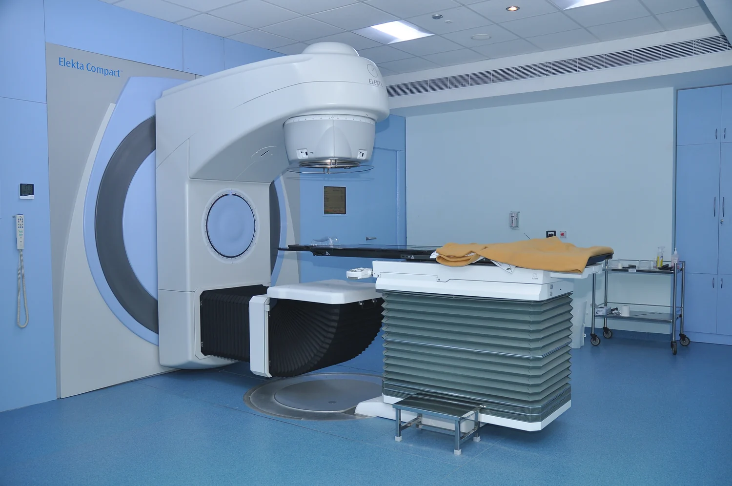
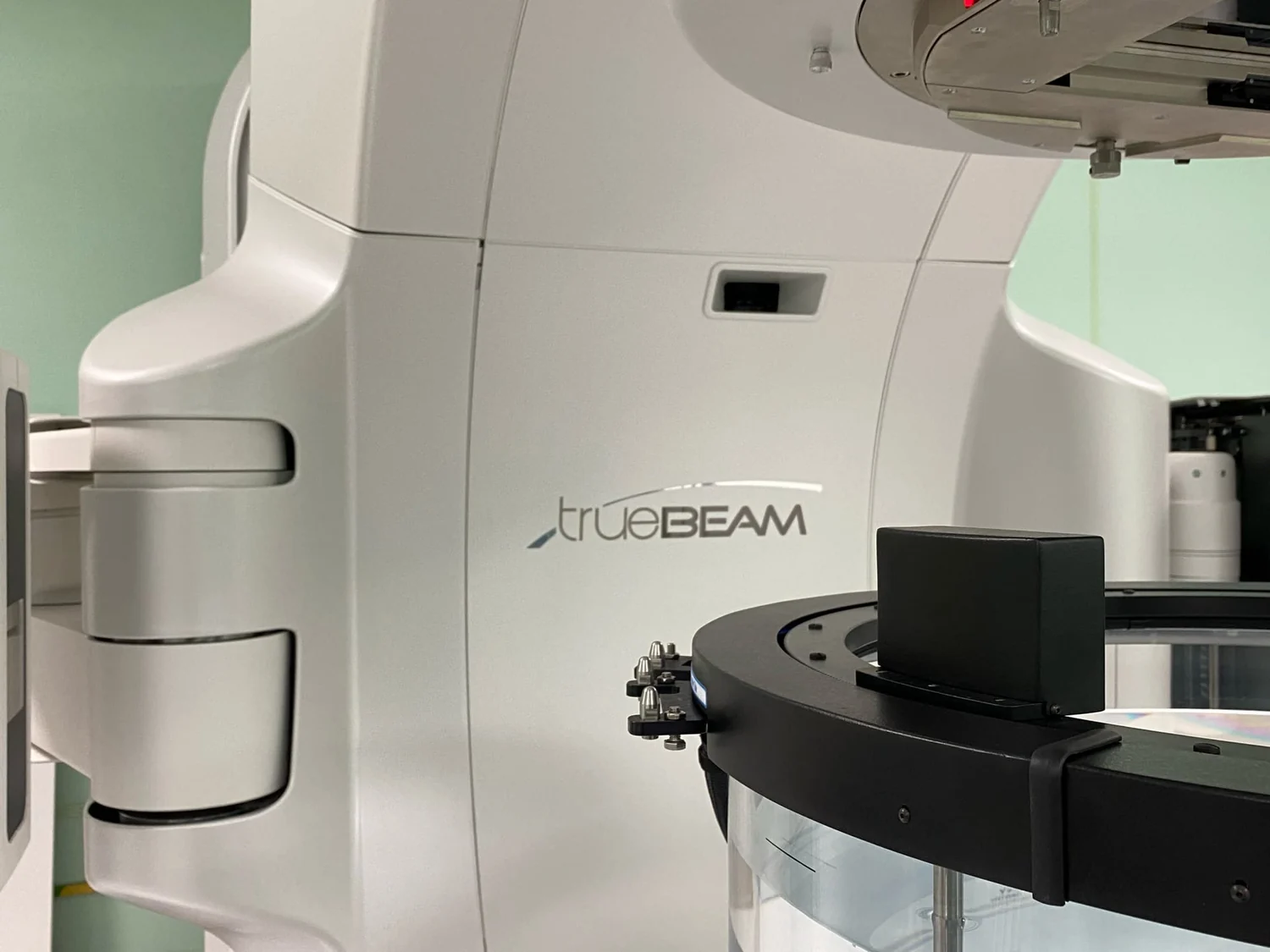
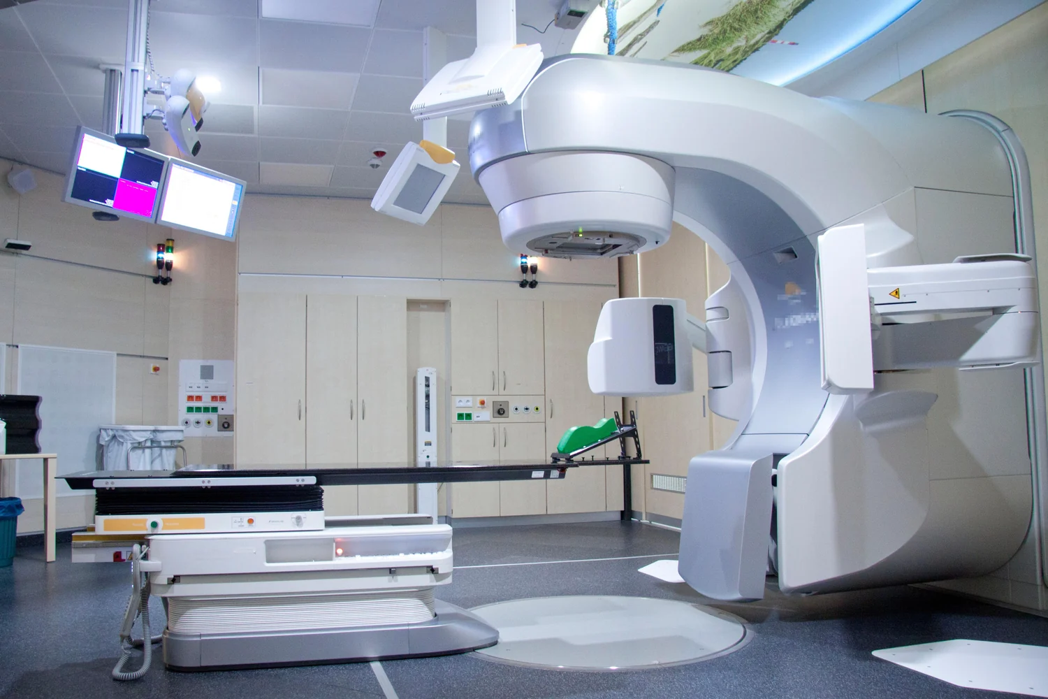
External beam radiation therapy comes from a machine aimed at a specific part of the body. For a lung tumor, for example, the treatment field is in the chest rather than the whole body [1]. Before treatment, imaging is used in a planning session called simulation; computers analyze those images to calculate a treatment area and beam paths.
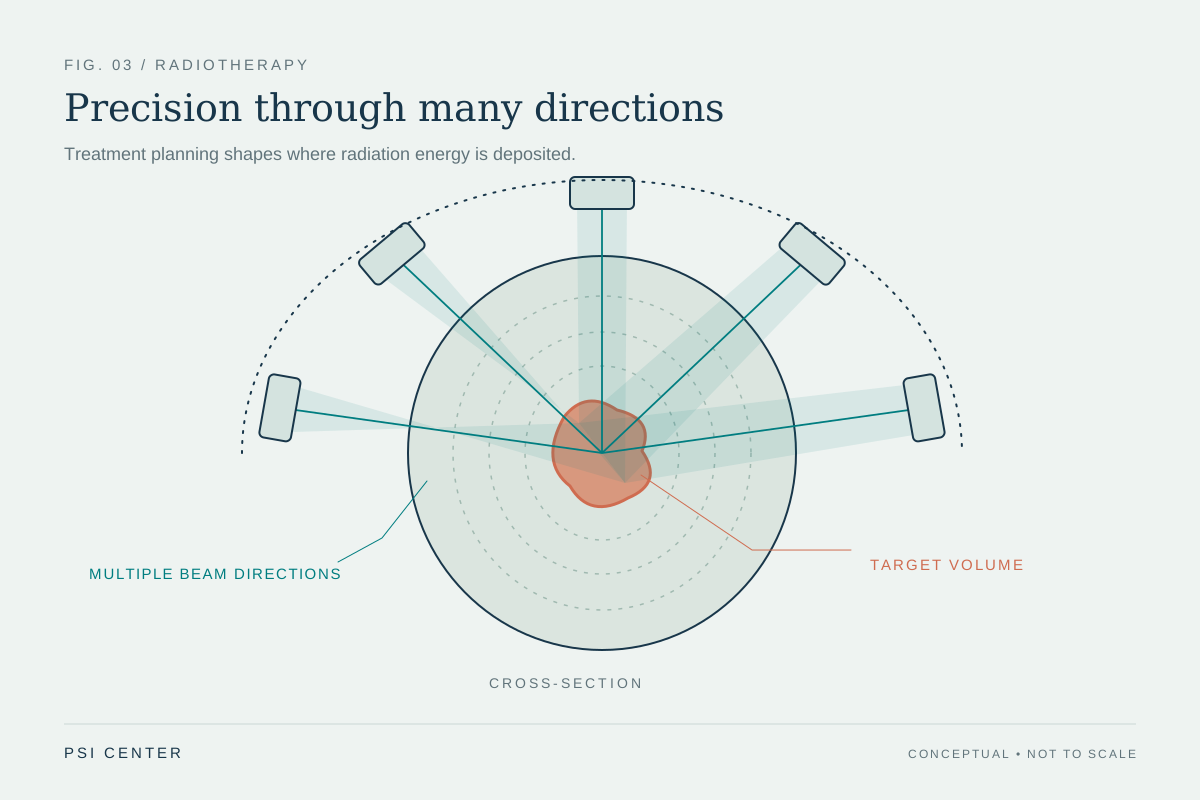
The geometry matters because a beam has to enter the body from somewhere. Three-dimensional conformal treatment uses beams from multiple directions shaped to fit the tumor, while intensity-modulated radiation therapy varies the strength of smaller beam segments to give different parts of the target different doses [1]. Each path crosses normal tissue, but the combined pattern can concentrate dose where beams converge.
Beam type changes where energy is deposited
Most radiation machines use photon beams. Photons can reach deep tumors, but they scatter some radiation along their path and continue beyond the tumor into normal tissue [1]. That means the treatment plan considers both the dose reaching the target and the dose delivered before and after it.
Protons have a different physical pattern: they can travel into the body and stop near the tumor, potentially reducing radiation to tissue beyond it. Whether that translates into a clinical advantage depends on the situation; trials compare proton and photon approaches, and machine size and cost limit proton availability [1]. Electrons travel only a limited distance in tissue, so their use is generally for tumors at or near the surface. No beam type eliminates the need to balance target coverage and nearby tissue exposure.
Position and motion affect precision
Even a carefully designed plan depends on placing the body consistently. Marks, molds, or masks can help reproduce the same position at each session [1]. Image-guided radiation therapy adds imaging during treatment sessions, allowing the team to detect changes in tumor size or location and adjust positioning or dose when needed [1]. These steps address the fact that anatomy is not a perfectly fixed diagram.
Breathing and digestion can move tumors, especially outside the brain. That motion makes precise targeting harder, which is one reason stereotactic body radiation therapy is often delivered in more than one dose rather than a single session [1]. Imaging and immobilization do not erase motion, but they help planners account for it and reduce the chance that the intended beam pattern misses its target.
Biology explains why treatment is often divided
Radiation damages DNA, which can stop cancer cells from dividing or lead them to die. The effect is not immediate: damaged cells can take days or weeks to die, and cell death may continue for weeks or months after treatment ends [2]. At the same time, radiation can affect nearby healthy cells, producing side effects. The physical plan therefore works alongside biological timing.
Many external beam courses use repeated sessions. Spacing treatments can give healthy cells time to recover and can make radiation more effective against cancer cells [1]. The schedule depends on the cancer, treatment goal, dose, and other factors; it is part of the plan rather than a uniform recipe. In this way, the balance is built from several decisions: where dose goes, how much reaches each region, how accurately the target is found, and how treatment is timed.
Explore the physics: an explanatory diagram
Sources & further reading
- External Beam Radiation Therapy National Cancer Institute
- Radiation Therapy to Treat Cancer National Cancer Institute
Prepared for the independent PSI Center reading library. About edition dates and source checking.
This explainer describes scientific principles. It does not assess individual treatment choices.