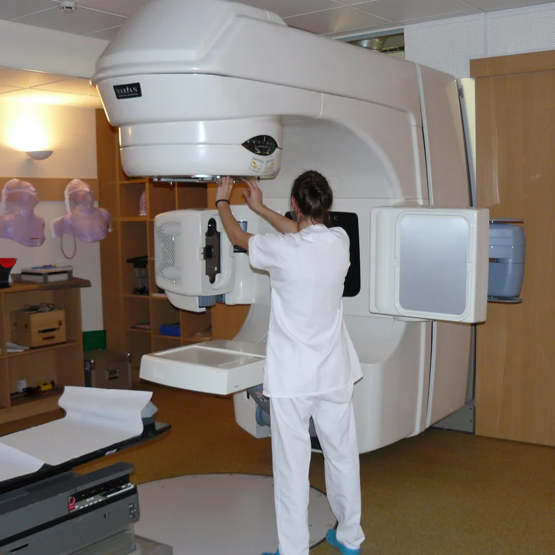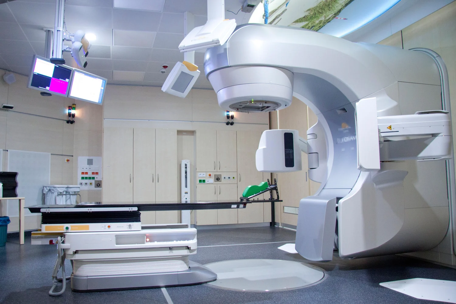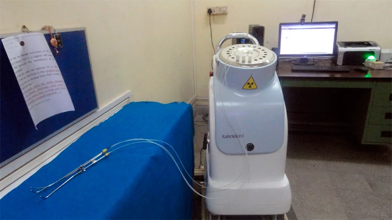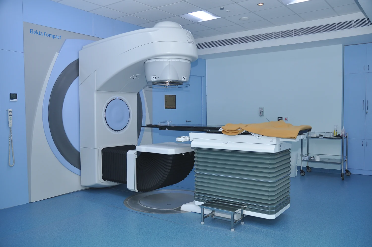
How can radiation enter the body from outside and still be aimed at a tumor inside? The answer is a chain of geometry and physics: images map the target, a plan chooses beam paths, and a machine delivers radiation from selected directions. Each beam travels through tissue, so the aim is to build up dose where paths converge while limiting exposure to nearby healthy structures.
Mapping the target before treatment
External beam radiation therapy is a local treatment: a machine directs radiation toward a selected body region rather than treating the whole body [1]. Before treatment, imaging is used to locate the tumor and nearby anatomy. A planning session, often called simulation, helps define the treatment area and the position the patient will use so the intended geometry can be reproduced.
The plan is computed to shape the beams around the tumor while sparing normal tissue as much as possible [1]. This is not a matter of the machine recognizing cancer by itself. Clinicians define the target and treatment plan from imaging and clinical information; the machine then delivers the planned beam arrangement with the patient positioned carefully.
Many paths can build one dose
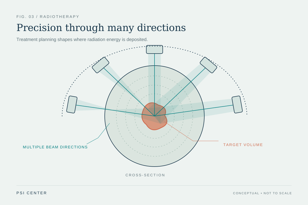
Imagine aiming several weak flashlights at the same spot on a wall. Along each route, light passes through other areas, but the overlap is brightest at the shared target. Radiation planning uses a related geometric principle: beams from several directions can combine dose in the tumor region, while each path contributes less dose to some surrounding tissue than a single direct route might.
Three-dimensional conformal therapy shapes beams to the tumor, while intensity-modulated radiation therapy (IMRT) uses many smaller beams and can vary their strength across regions. Image-guided radiation therapy adds scans during treatment sessions so changes in target position or size can inform adjustments [1]. These are ways to refine delivery, not guarantees that nearby tissue receives no radiation.
Different particles, different paths
Most external beam machines use photon beams. Photons can reach tumors deep in the body, but they scatter some radiation along their path and do not stop at the tumor; normal tissue beyond the target can also receive exposure [1]. This physical behavior is one reason planning considers both the entrance and exit paths of a beam.
Protons are charged particles that can be planned to stop near the target, potentially reducing dose to some normal tissue beyond it. Whether that advantage improves outcomes compared with photon treatment is a clinical question; the source describes trials comparing the approaches and notes that cost and machine size limit proton use [1]. Electrons, with shorter travel through tissue, are generally used for tumors at or near the surface [1].
Dose, time, and tissue response
Radiation damages the DNA of cells. Cancer cells with damage they cannot repair stop dividing or die, but this response takes time: cell death can continue for weeks or months after treatment ends [2]. Radiation can also affect nearby healthy cells, which is why treatment plans balance tumor control with limiting unintended exposure [2].
A course is often divided into multiple sessions, or fractions. Spacing treatments gives healthy cells time to recover and can make radiation more effective; schedules vary with the cancer, goal, dose, and plan [1] [2]. External beam treatment does not make the patient radioactive [1]. The machine’s movement may be visible and audible, but the essential action is an energy delivery guided by imaging, position, and a calculated dose pattern.
Explore the physics: an explanatory diagram
Sources & further reading
- External Beam Radiation Therapy National Cancer Institute
- Radiation Therapy to Treat Cancer National Cancer Institute
Prepared for the independent PSI Center reading library. About edition dates and source checking.
This explainer describes scientific principles. It does not assess individual treatment choices.