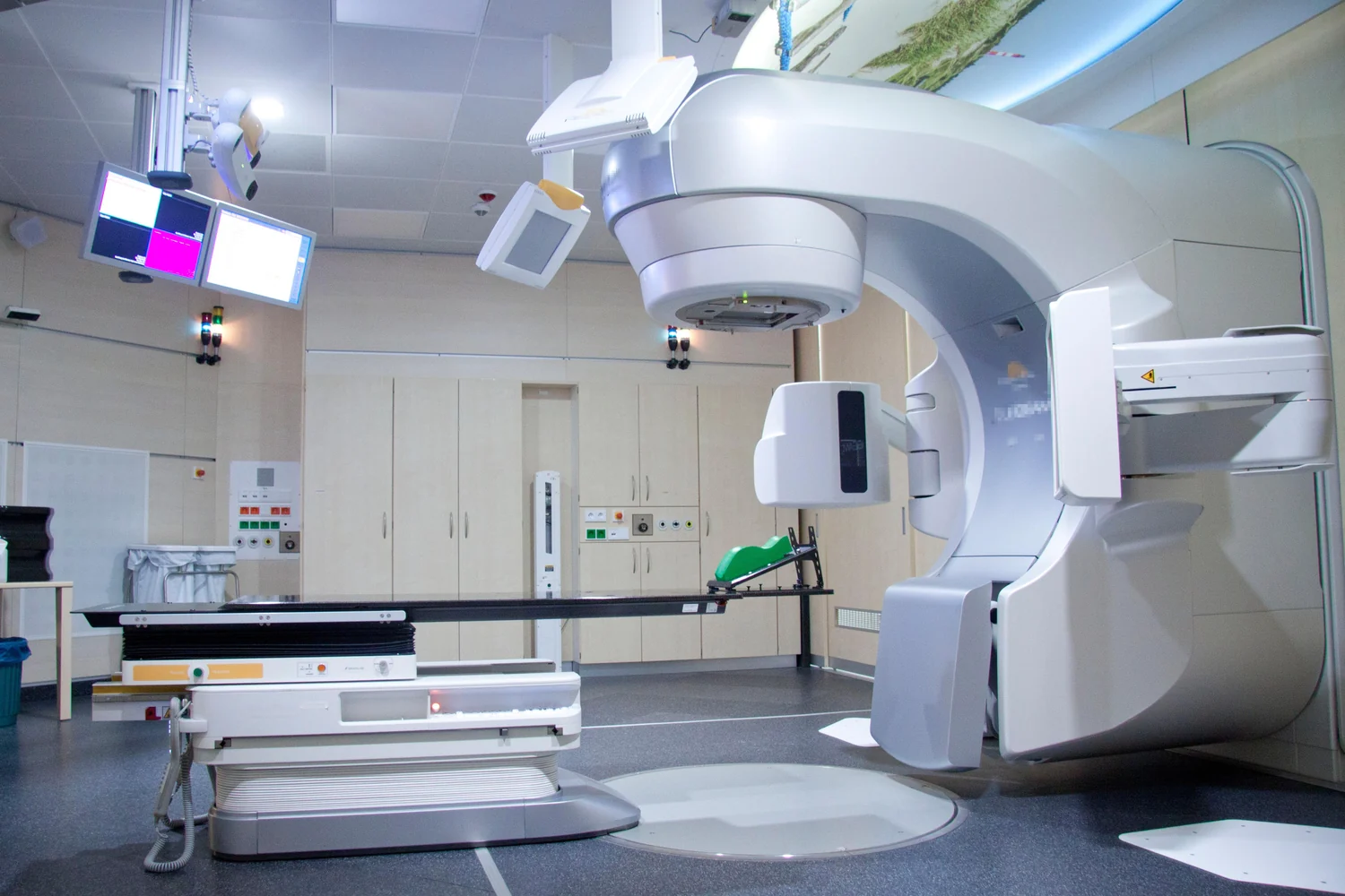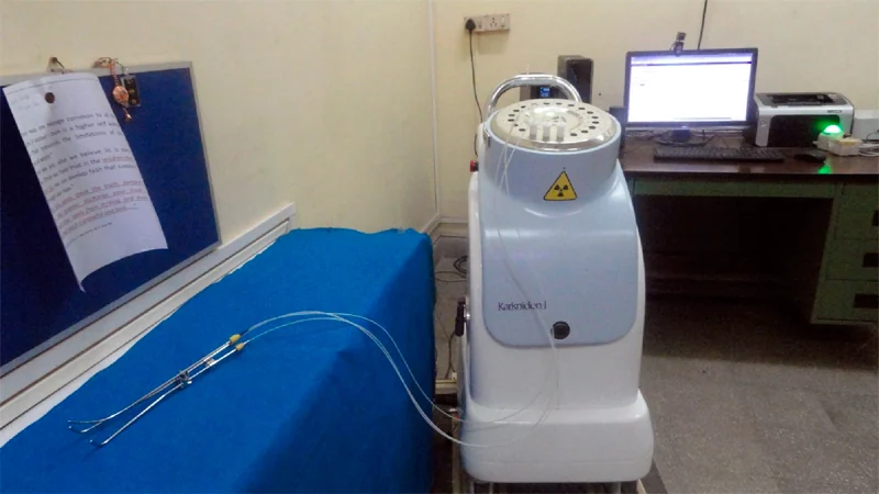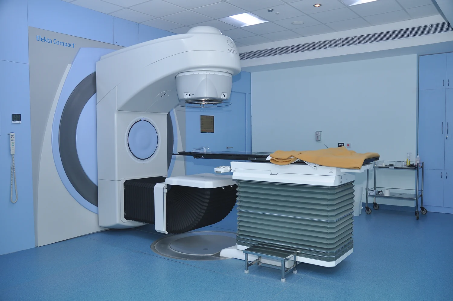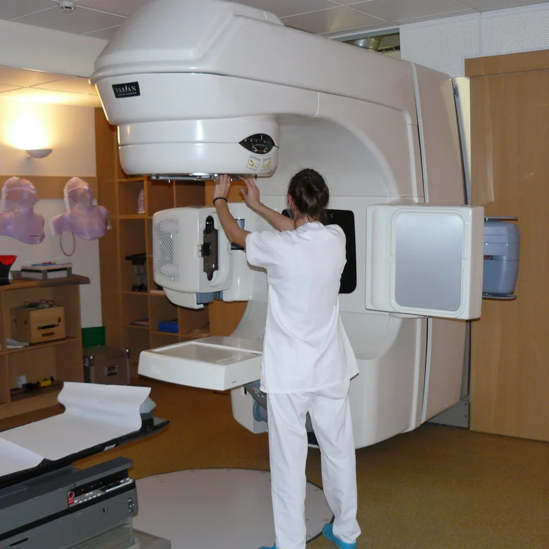
How can radiation reach a tumor deep inside the body without sending all of its energy through the same strip of healthy tissue? One answer is to aim beams from several directions. Each path contributes dose as it travels, while their overlap can be arranged at the target. Imaging and computation help shape those paths around a three-dimensional problem: where the tumor lies, how it is shaped, and what normal structures sit nearby.
Every path has a cost
External beam radiation therapy is a local treatment: a machine aims radiation at a particular region rather than treating the whole body [1]. But a deep target cannot be reached without radiation crossing tissue on the way in. For photon beams, some radiation is scattered along the path, and the beam continues beyond the tumor into tissue behind it. A single direction can therefore expose a corridor of normal tissue as well as the intended target [1].
Imagine shining a flashlight through a layered fruit to reach a seed in the middle. Light must cross the peel and flesh before it reaches the seed, and it continues beyond it. Radiation is not visible light, and the tissues are not fruit, but the geometry offers a useful distinction: reaching an interior point means accounting for what lies before and after it. Treatment planning is a way to manage that geometry.
Overlap can make a target stand out
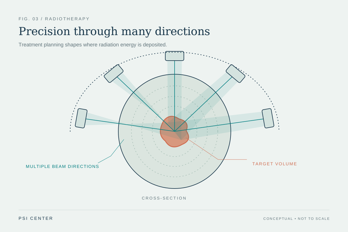
When beams enter from different directions, the paths through normal tissue are distributed around the target. The plan can arrange for their contributions to overlap at the tumor, where the prescribed dose is intended, while reducing the contribution from any single entry path. In stereotactic radiosurgery, for instance, many small beams are aimed from different directions; each has little effect on the tissue it passes through, while the focused dose is delivered where they converge [1].
The same principle appears in three-dimensional conformal radiation therapy, which uses imaging to map the tumor and computer calculations to design beams that conform to its shape. Intensity-modulated radiation therapy takes the idea further with many smaller beams and changes in beam strength across regions, allowing higher dose to selected parts of the tumor [1]. Several directions are not a guarantee that every nearby structure is spared; they provide more ways to distribute and shape dose.
A plan is a calculated compromise
A treatment team needs a map of the target and surrounding anatomy. CT, MRI, and PET images may be used in simulation, the planning process that defines the treatment area. A computer then analyzes the images and calculates beam paths and dose distributions [1]. This is a complex system with many interacting variables, so computational models can simulate how changing inputs affect predicted outputs [2].
A model is not a direct measurement of what every cell will experience. It is a mathematical representation built from assumptions and available information. Mechanistic models use known physical principles, while data-driven models identify patterns in datasets; some combine both approaches [2]. The value of computation here is that planners can compare many candidate arrangements in a virtual setting before choosing a workable treatment plan.
Position and movement matter too
Even a carefully shaped plan depends on placing the patient and target where expected. Marks, body molds, or masks can help reproduce the same position at each session. Some methods also image during treatment and use repeated scans to detect changes in tumor size or location; image-guided radiation therapy can support adjustments to position or dose when needed [1]. A moving target, such as one affected by breathing, poses a different challenge from a small, still target in the brain.
The several-direction strategy is therefore a way of controlling where dose accumulates, guided by images and calculations and constrained by anatomy and motion. It does not make radiation selective in the sense of affecting only tumor cells: normal tissue along beam paths still matters. The plan’s job is to deliver the prescribed dose to the intended region while sparing surrounding tissue as much as the technique and clinical situation allow [1].
Explore the physics: an explanatory diagram
Sources & further reading
- External Beam Radiation Therapy National Cancer Institute
- Computational Modeling NIBIB
Prepared for the independent PSI Center reading library. About edition dates and source checking.
This explainer describes scientific principles. It does not assess individual treatment choices.