What changes when a radiation beam is divided into many smaller regions with different strengths? In intensity-modulated radiation therapy, or IMRT, the treatment team changes how much radiation is delivered from different parts of the beam. The aim is to fit the planned dose to a tumor's shape while limiting exposure to nearby healthy tissue.
Shaping dose through the beam
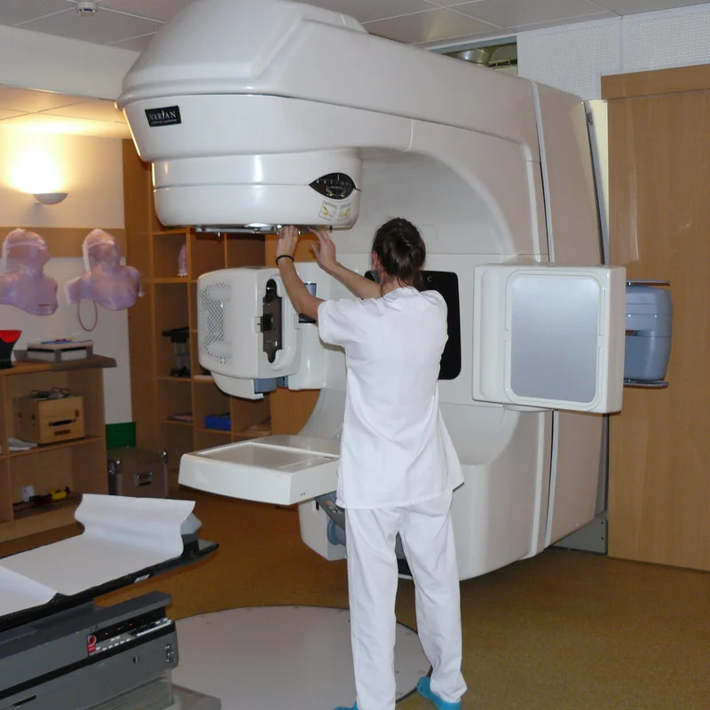
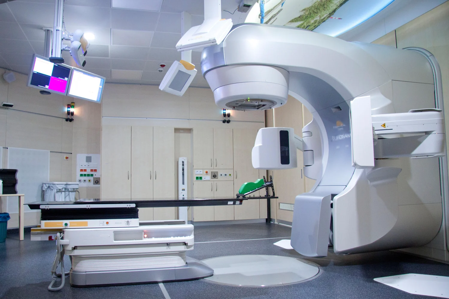
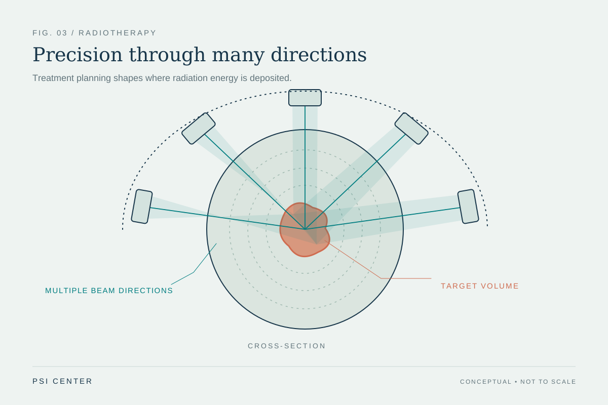
External-beam radiation therapy uses a machine to aim radiation at a specific part of the body; it is a local treatment [1]. IMRT is a form of three-dimensional conformal radiation therapy. Like that approach, it directs beams toward a tumor from several directions, but it uses many more, smaller beams and varies their strength across the beam [1].
Picture a landscape whose hills and valleys represent how much radiation reaches each point. A uniform beam would contribute a broadly similar intensity across its field. IMRT can vary intensity across small regions, allowing the combined dose from multiple directions to be shaped more closely around the target. The variation is planned, not improvised: computers analyze images of the tumor to calculate a treatment path and dose distribution [1].
Why use several directions?
Each beam travels through tissue on its way to the tumor. With photon beams, radiation is scattered along the path, and the beam continues beyond the tumor into normal tissue [1]. A single entry direction may therefore expose healthy structures in front of and behind the target. When beams enter from multiple directions, their contributions can overlap at the tumor while individual paths carry smaller portions of the total planned dose.
This is a geometric strategy, not a way to make radiation vanish outside the target. The dose from each direction accumulates, and sensitive nearby structures still constrain the plan. The goal shared by external-beam techniques is to deliver the highest prescribed dose to the tumor while sparing surrounding normal tissue [1]. In a complicated target shape, changing intensity across a beam gives planners another way to distribute that accumulated dose.
Images and models guide the plan
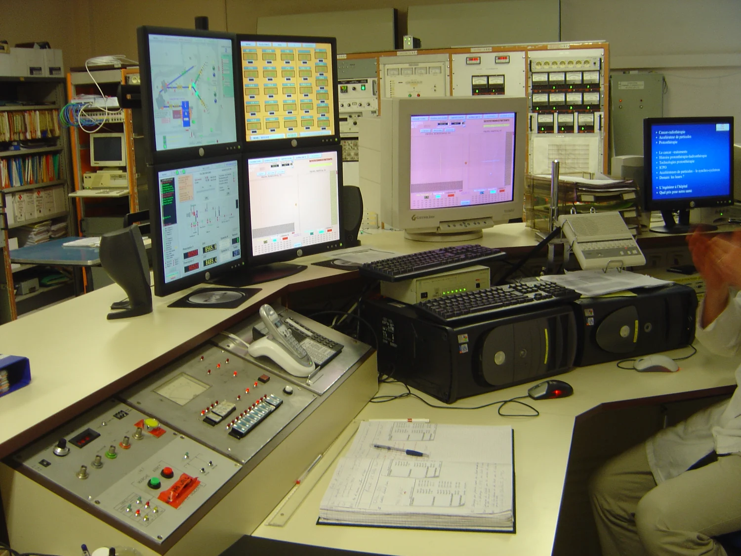
Before treatment, images such as CT, MRI, or PET can be used to plan the treatment area and shape the radiation beams [1]. A computer model represents the patient and treatment geometry in a form that can be used to calculate what different beam arrangements would do. Computational models generally simulate complex systems using mathematics, physics, and computer science; they can incorporate established mechanisms, observed data, or both [2].
For radiation planning, the calculation is a prediction about dose under specified conditions. It depends on the images, the representation of tissue, the beam descriptions, and the assumptions built into the model. Modeling can compare candidate arrangements efficiently, but a simulated result is not itself a measurement of what happens in a patient's body. Planning and delivery procedures must translate the calculated arrangement into a reproducible treatment.
Precision depends on the whole process
IMRT describes how beam intensity is modulated; image-guided radiation therapy describes adding scans during treatment sessions to detect changes in tumor size or position and allow adjustments if needed [1]. They are related but distinct ideas. A precisely shaped plan still depends on positioning the patient consistently and accurately identifying the target. Some tumors can also move with ordinary body motion, such as breathing or digestion, which complicates targeting [1].
The model's role is to help calculate a distribution, not to promise a particular outcome. Computational modeling can simulate systems at different scales, and its predictions depend on the variables and rules included [2]. For a reader, the central distinction is between shaping the planned dose and guaranteeing that every cell receives an identical amount. IMRT changes the distribution of radiation across directions and beam regions so that clinicians can pursue a target dose while accounting for the healthy tissues around it.
Explore the physics: an explanatory diagram
Sources & further reading
- External Beam Radiation Therapy National Cancer Institute
- Computational Modeling NIBIB
Prepared for the independent PSI Center reading library. About edition dates and source checking.
This explainer describes scientific principles. It does not assess individual treatment choices.