If a tumor shifts as the lungs expand and relax, where should a radiation beam be aimed? External beam therapy is planned to deliver a prescribed dose to a specific region while limiting exposure to nearby healthy tissue. Breathing makes that target a moving one. The planning problem is therefore not only to map the tumor, but also to understand how its position changes and how the treatment can account for that motion.
A moving target changes the geometry
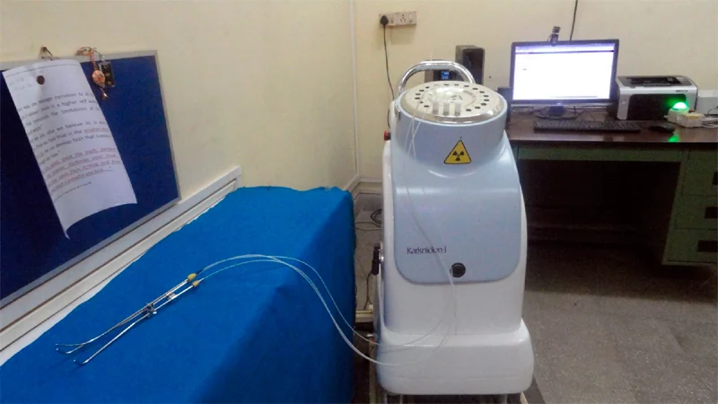
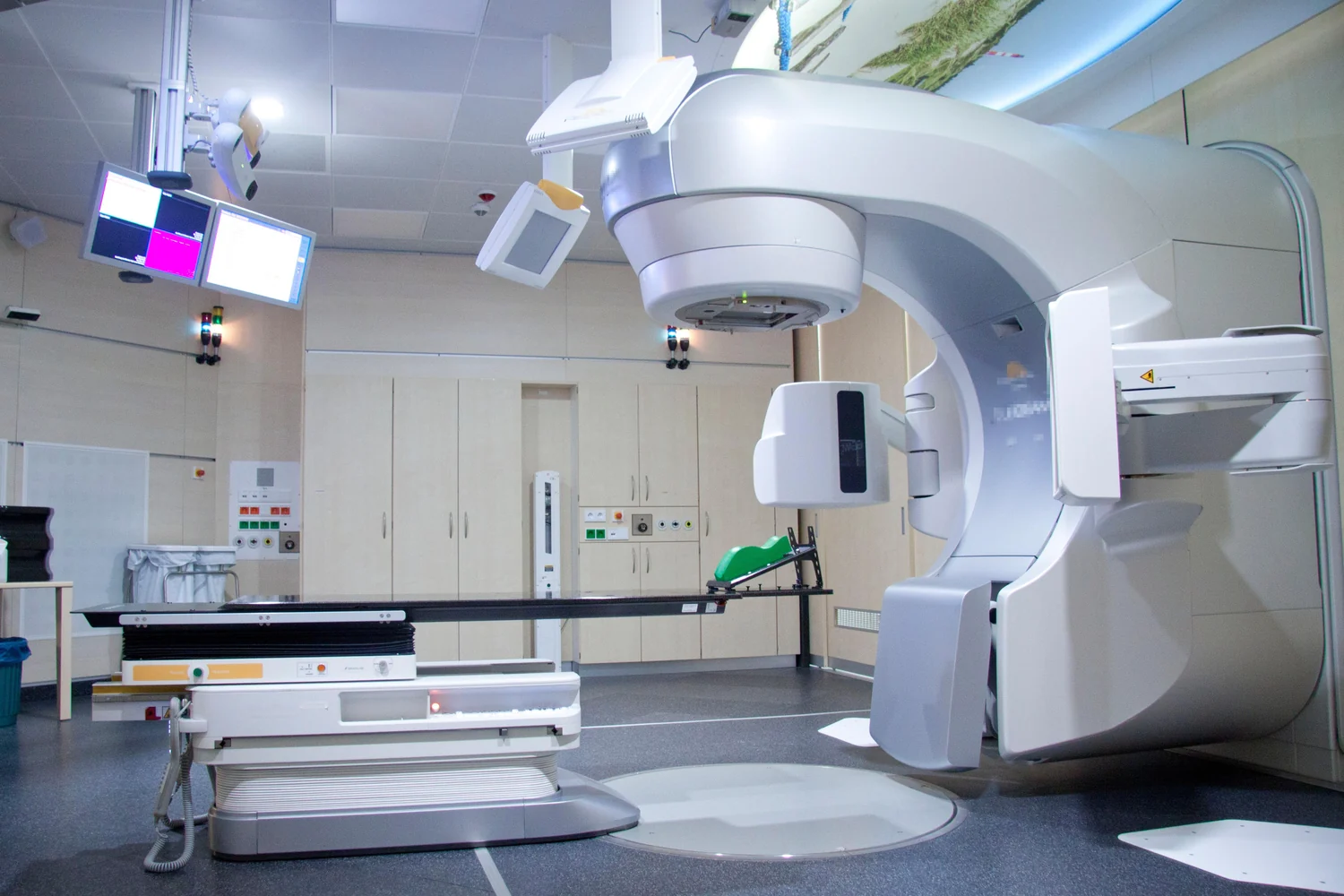
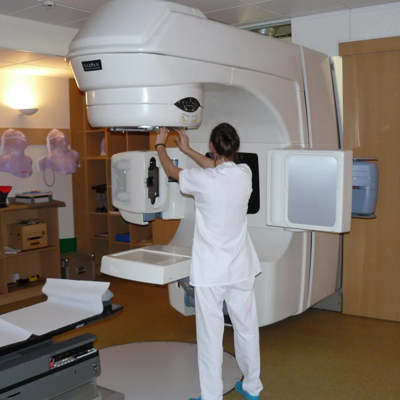
External beam radiation therapy uses a machine to direct radiation at a treatment area. It is a local treatment: the goal is to treat a specific part of the body. Planning uses imaging to define the tumor and nearby anatomy, then a computer calculates a treatment path intended to deliver the prescribed dose while sparing surrounding normal tissue [1].
For a tumor in the lung, breathing can shift the target relative to the machine and nearby organs. A plan based on a single position may not represent every moment during treatment. The size and direction of motion matter because a beam that is accurately placed at one instant may intersect a different volume as the body moves. This makes motion part of the geometry that planning must consider.
Images capture position; models explore change
Radiation planning often uses CT, MRI, or PET images to define a treatment area. During image-guided radiation therapy, scans can also be taken during treatment sessions, and computers can detect changes in tumor size or location so positioning or dose can be adjusted if needed [1]. Repeated imaging helps account for changes over time, though an image is still a measurement of anatomy at a particular moment.
Computational modeling uses mathematics, physics, and computer science to simulate complex systems under different conditions [2]. A model can represent variables that affect a system and predict outputs for scenarios that are difficult to measure directly. In a radiation-planning context, this general approach helps explain why teams may examine how a target’s position varies rather than treating the anatomy as motionless. The model’s usefulness depends on the information and assumptions built into it.
Why a small shift can matter
Picture drawing a narrow circle around a moving point on a map. If the point drifts while the circle remains fixed, some of its path will fall outside the boundary. Radiation beams create a related planning challenge: treatment must cover the intended target despite motion, while limiting dose to healthy tissue nearby. The more tightly a treatment is shaped around a target, the more consequential uncertainty in its position can become.
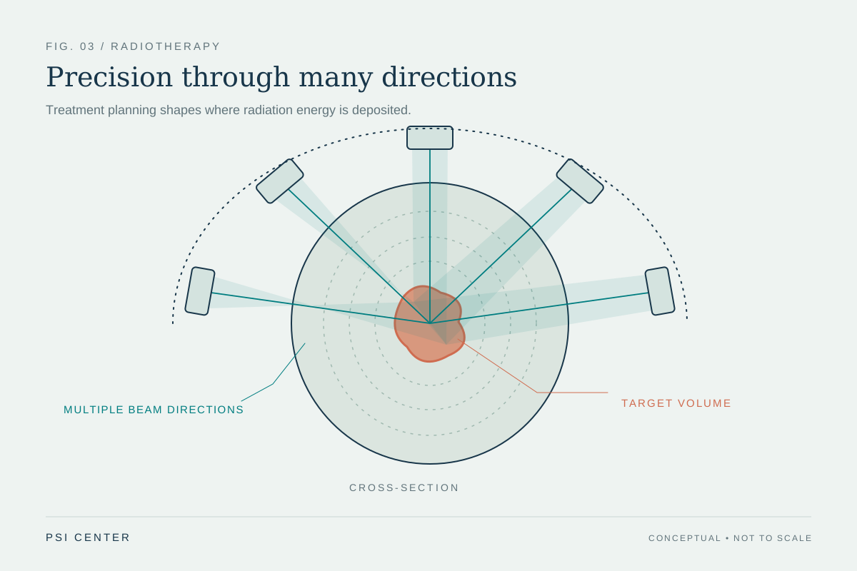
External beam methods use different strategies to shape delivery. Three-dimensional conformal radiation uses beams from multiple directions, and intensity-modulated radiation therapy varies beam strength across smaller segments. Image-guided radiation adds imaging during sessions to detect changes and support adjustments [1]. These approaches share the goal of precise delivery, but their details and suitability depend on the treatment situation.
Precision depends on information and assumptions
A computational model is a representation, not the moving body itself. Mechanistic models use established principles about how a system works; data-driven models draw on patterns in datasets; some models combine both approaches [2]. For motion questions, the model’s output is only as useful as its inputs and the way those inputs capture the relevant changes. A predicted path should not be confused with a direct observation at every moment.
That is why planning links imaging, calculation, positioning, and verification. External beam treatments use simulation to determine the treatment area, and image-guided approaches can repeat scans during sessions to identify shifts [1]. Breathing introduces a changing coordinate system: the target’s location moves relative to the body surface and treatment machine. Understanding that movement helps the treatment team reason about coverage and nearby tissue, turning a static picture into a plan that acknowledges motion.
Explore the physics: an explanatory diagram
Sources & further reading
- External Beam Radiation Therapy National Cancer Institute
- Computational Modeling NIBIB
Prepared for the independent PSI Center reading library. About edition dates and source checking.
This explainer describes scientific principles. It does not assess individual treatment choices.