How can one tiny inner-ear structure distinguish a high whistle from a low drumbeat? The cochlea does not need a separate chamber for every note. Instead, sound vibrations travel through its fluid and create a wave along an elastic partition. Different frequencies peak at different places, giving the brain an ordered map of pitch.
Sound arrives as vibration
Air pressure changes from a sound wave enter the ear canal and vibrate the eardrum. Three small middle-ear bones—the malleus, incus, and stapes—pass and amplify those vibrations toward the fluid-filled cochlea [1]. The chain matters: airborne motion becomes mechanical motion, then fluid motion inside the inner ear.
The cochlea is shaped like a spiral, but its essential sorting mechanism is a long elastic partition called the basilar membrane. It runs through the cochlea and supports sensory hair cells. When incoming vibrations ripple the cochlear fluid, they launch a traveling wave along that membrane [1].
A wave has a preferred place to peak
The basilar membrane’s response varies along its length. A high-pitched sound produces its strongest motion near the wide end of the cochlea, while lower-pitched sounds peak closer to the center [1]. In effect, pitch becomes position: a tone’s frequency determines where the traveling wave has its greatest effect.
Picture a long spring or rope fixed at one end and struck with different rhythms. The details are not identical to the cochlea, but the thought experiment captures an important point: a material system can respond differently at different locations depending on the frequency of the incoming vibration. The cochlea’s geometry and mechanics create a biological frequency map.
Hair cells turn motion into signals
Hair cells sit on the basilar membrane and move with its wave. Each cell carries tiny projections called stereocilia. As those projections bend against an overlying structure, pore-like channels at their tips open; chemicals enter the cell and trigger an electrical signal [1]. The ear thus converts a mechanical event into a message the nervous system can carry.
The location of active hair cells gives the signal information about pitch, while the timing and pattern of activity carry other features of sound. The auditory nerve transmits these electrical signals to the brain, which interprets them as sounds we recognize [1]. The cochlea performs an early sorting step; perception depends on later neural processing too.
The map is useful, but hearing is a chain
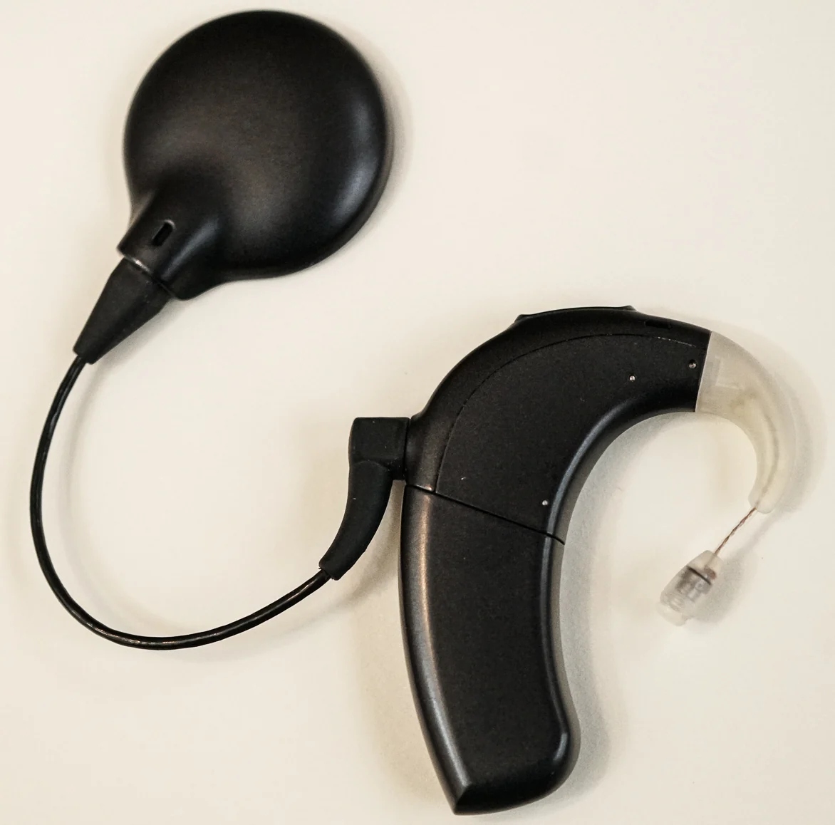
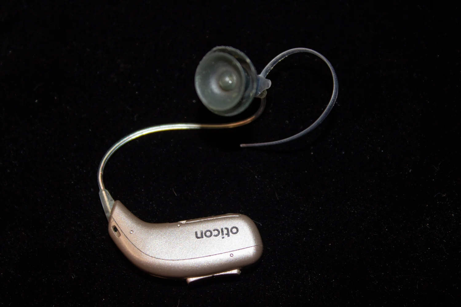
Frequency separation helps explain why a hearing aid can be programmed to amplify some pitches more than others. Digital hearing aids encode sound numerically, allowing adjustments by frequency and, in some devices, by sound direction [2]. Yet amplification cannot guarantee that damaged hair cells will turn larger vibrations into neural signals; there are practical limits when inner-ear damage is extensive [2].
That distinction shows why the cochlea’s map is only one part of hearing. Sound must pass through the outer and middle ear, move cochlear fluid, stimulate sensory cells, and travel along the auditory nerve before the brain recognizes a voice or melody. A pitch has a physical frequency, but hearing it depends on a sequence of functioning transformations.
Sources & further reading
- How Do We Hear? NIDCD
- Hearing Aids NIDCD
Prepared for the independent PSI Center reading library. About edition dates and source checking.
This explainer describes scientific principles. It does not assess individual treatment choices.