How can a clinician inspect layers beneath a tissue surface without cutting into it? Optical coherence tomography, or OCT, answers with light, while ultrasound answers with sound. Both can turn returning signals into cross-sectional images, which is why they are often compared. But the analogy has limits: light and sound travel, scatter, and interact with tissue differently, so the two methods suit different depths and settings.
A shared idea: send a wave, read what returns
Picture a short pulse entering tissue and encountering a boundary between two materials. Some of its energy continues onward; some returns toward the instrument. If the device can detect that returning signal and estimate where it came from, it can build a map of internal layers. Ultrasound does this with high-frequency sound waves. A transducer sends sound into the body and detects echoes reflected at tissue boundaries. Knowing the speed of sound in tissue and measuring an echo’s return time lets the scanner estimate the boundary’s distance from the probe. Many such measurements form an image. [2]
OCT uses a related imaging logic, but its probe is infrared light rather than sound. Light entering tissue is absorbed, scattered, or reflected; the returning light carries information about structures below the surface. OCT is used for detailed views within the retina, and cardiologists use it to help diagnose coronary artery disease. The comparison is therefore useful at the level of the question both methods ask: what structures lie along a path, and what signal comes back from them? [1]
The clocks work differently
In conventional ultrasound, timing is central. The scanner records how long an echo takes to return, then uses an assumed sound speed to estimate depth. A familiar example is listening for an echo in a canyon: a longer delay suggests a more distant wall. In a medical scan the distances are far smaller, but the principle is similar. Boundaries matter because changes in tissue properties can send part of a sound wave back toward the transducer. [2]
OCT also needs information about where returning light originated, but its measurement is not simply a sound-style stopwatch. Its name refers to optical coherence: the method uses light-wave interference to distinguish returns associated with different depths. That distinction helps explain why an OCT image is not just an ultrasound picture drawn with a different color palette. It is a reconstruction based on a different kind of signal and a different measurement strategy. Both methods make depth visible, but the physical route to that depth is not interchangeable. [1]
Why a useful analogy has limits
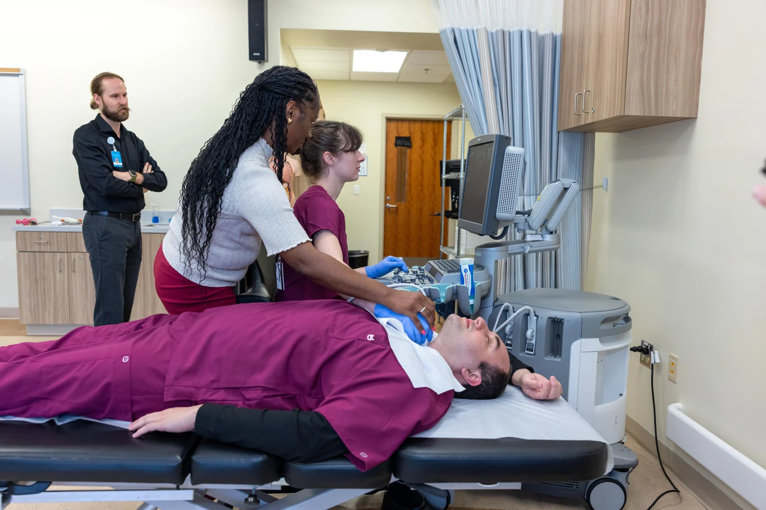
Sound can travel through many soft tissues and return as echoes from boundaries, allowing ultrasound to image organs, muscles, blood vessels, and other structures. But ultrasound is poor at imaging bone and tissues containing air, such as the lungs, because those materials interfere with sound transmission. The practical image also depends on keeping air pockets out of the path between the probe and skin, which is why gel is applied during a scan. [2]
Light is strongly scattered in tissue, so OCT is particularly suited to detailed views over limited subsurface distances rather than a general scan through the whole body. NIBIB describes OCT as obtaining subsurface images of structures just below the skin and notes its uses in the retina and coronary arteries. The broader family of optical imaging methods can reveal tissue properties through how different tissues absorb and scatter light, but those methods do not all share OCT’s specific design or depth. [1]
Two instruments, different questions
Imagine comparing a cross-section of the eye with an image of a developing fetus. OCT’s detailed retinal view makes it useful in ophthalmology, while ultrasound’s ability to image internal organs supports pregnancy monitoring and many other examinations. In either case, the clinician is not simply seeing a photograph. The image is inferred from how waves interact with tissue and from assumptions built into the instrument’s reconstruction. [1] [2]
The comparison becomes most helpful when it prompts a better question: which wave can reach the structure of interest, and what property of the returning signal can reveal it? Ultrasound can also make functional maps of blood flow or tissue stiffness, while optical imaging can measure changes related to soft-tissue properties. Similarities in image shape do not imply identical information. The shared idea is wave-based probing; the useful choice depends on the tissue, the needed detail, and the signal the method can reliably collect. [1] [2]
Explore the physics: an explanatory diagram

Sources & further reading
- Optical Imaging NIBIB
- Ultrasound NIBIB
Prepared for the independent PSI Center reading library. About edition dates and source checking.
This explainer describes scientific principles. It does not assess individual treatment choices.