Why can a beam of light show a crisp image of the retina but struggle to map tissue deeper in the body? Light does not travel through flesh like a pencil line through clear glass. Its photons are absorbed, redirected, or returned, and each tissue imposes a different mix of those effects. The resulting depth limit is not simply a weakness of light; it shapes which optical method can answer a particular question.
A photon’s path is constantly interrupted
When light enters tissue, molecules and structures can absorb some of its energy. Other photons scatter: they change direction after interacting with cells, fibers, or other microscopic features. Some light is reflected back toward a detector, while some may be emitted after the tissue absorbs energy. Optical imaging turns these interactions into information, but the same events that carry information also blur or weaken it. [1]
Imagine shining a flashlight through a clear pane and then through a milky glass. In the pane, many rays stay on course; in the milk glass, they spread into many directions. Tissue is more complicated still, because its composition varies from place to place. A detector may receive plenty of light without being able to tell where it came from. Thus, “how much light gets through” and “how clearly a feature can be located” are different questions.
Wavelength changes the trade-off
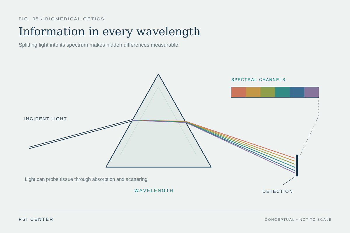
Different wavelengths interact with tissue differently. Near-infrared light is useful for diffuse optical methods that estimate properties such as total hemoglobin concentration and blood oxygen saturation in soft tissue. Yet a broad, scattered glow does not automatically produce a sharply localized image. The method can be valuable precisely because it measures how light has been altered across a volume, rather than trying to preserve a straight path to one tiny feature. [1]
At another scale, optical coherence tomography (OCT) uses infrared light to create subsurface cross-sections. Ophthalmologists use it to image structures within the retina. OCT’s comparison with ultrasound is instructive: the two methods both infer internal boundaries from returned signals, but one uses light and the other uses sound waves. Their different interactions with tissue make them suitable for different imaging settings. [1]
A boundary can help—or block—the view
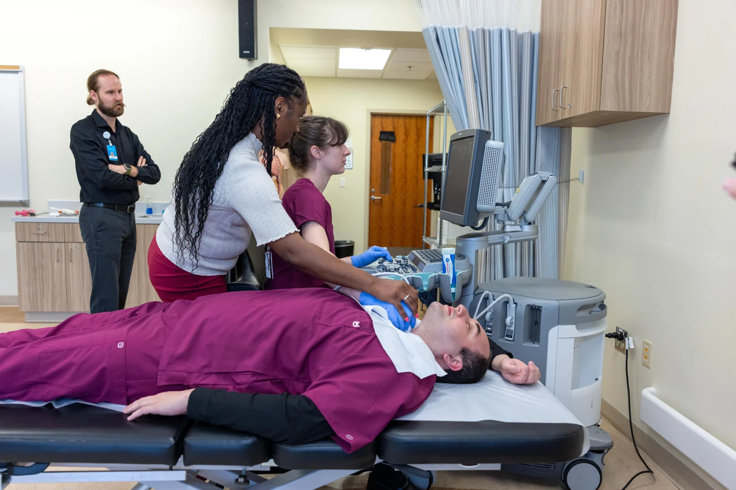
A tissue boundary can redirect or reflect a wave, making a change in structure visible. Diagnostic ultrasound uses echoes: a transducer sends sound into the body and detects reflections from boundaries such as fluid next to soft tissue. The time taken by an echo to return helps a scanner estimate the boundary’s distance. This is not the same mechanism as optical imaging, but it offers a useful comparison: image depth depends on how a signal travels and returns through its surroundings. [2]
Ultrasound, too, has obstacles. It is not good at imaging bones or air-containing tissues, because those materials interfere with the sound path. Light’s obstacles differ: absorption and scattering can scramble its direction and reduce the useful signal. A method’s limitation is therefore tied to the physical contrast it uses, not to a universal ranking in which one kind of wave always sees deeper.
Sometimes the signal takes a different route
Photoacoustic imaging turns absorbed light into sound. Laser pulses are delivered to tissue; absorbing regions warm slightly and generate ultrasound waves that can be detected by a transducer. This hybrid approach can create high-resolution images deeper in the body than purely optical imaging can readily provide. It does not mean light has suddenly stopped scattering; rather, the detected signal changes from light to sound after absorption. [1]
The choice of method follows the question. OCT can reveal fine subsurface structure in the retina; diffuse optical imaging can estimate oxygenation in soft tissue; photoacoustic imaging can use absorption to map features such as blood vessels. Ultrasound offers a different set of contrasts and limits. “Seeing depth” is not one property of a device: it emerges from wavelength, tissue, signal conversion, and the detector’s ability to distinguish a meaningful return from a cloud of scattered energy.
Explore the physics: an explanatory diagram
Sources & further reading
- Optical Imaging NIBIB
- Ultrasound NIBIB
Prepared for the independent PSI Center reading library. About edition dates and source checking.
This explainer describes scientific principles. It does not assess individual treatment choices.