What can a pulse of near-infrared light reveal about tissue that a camera cannot see? As light travels through skin and soft tissue, blood and other molecules absorb some wavelengths while tissue scatters others. Measuring the returning or transmitted light can therefore provide clues about what lies below the surface. Those clues are indirect: the instrument measures photons, then estimates biological properties from how they were altered.
Light takes a complicated path
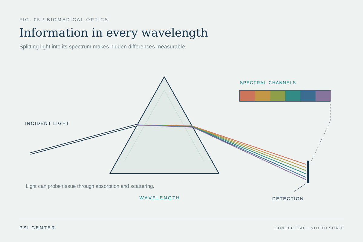
Near-infrared light sits just beyond visible red in the electromagnetic spectrum. It can penetrate soft tissue more deeply than many visible wavelengths, although it does not travel in a straight, unobstructed line. Molecules absorb particular portions of the spectrum, while structures such as cells and fibers scatter light in many directions. Optical imaging uses these interactions—absorption, scattering, reflection, and emission—to study biological material [1].
A simple picture is a flashlight shining through fog: the light that emerges has been weakened and redirected. Tissue is more complex than fog because its composition varies from point to point, but the analogy captures why measurement is difficult. A detector may receive photons that took different paths and crossed different mixtures of tissue. The amount of light alone rarely identifies a single cause.
Blood leaves an optical signature
Hemoglobin, the oxygen-carrying protein in red blood cells, absorbs light differently depending on whether it is carrying oxygen. A pulse oximeter uses red and near-infrared light passing through a fingertip and compares the changing absorption signals to estimate blood oxygen saturation [2]. The pulse component helps distinguish changing arterial blood volume from more constant absorption by surrounding tissue.
Diffuse optical imaging applies related principles across larger regions. Researchers can use near-infrared measurements to estimate total hemoglobin concentration and blood oxygen saturation in soft tissue [1]. These estimates can help study blood supply or tissue function, but they are not direct photographs of oxygen molecules. The device interprets an optical signal through assumptions about light paths and tissue properties.
Why the estimate can shift
Consider a finger sensor during movement. If the finger shifts, the path between light source and detector changes; the sensor may record motion artifact rather than a genuine oxygen change. Poor blood flow can weaken the pulse signal. Nail polish can alter the light that reaches the detector, and skin pigment also absorbs red and near-infrared light. These effects can bias readings, including a tendency for pulse oximeters to overestimate oxygen saturation in people with darker skin tones [2].
This example illustrates a general measurement principle: a sensor observes an interaction, not the underlying quantity in isolation. The same tissue can produce different signals when probe pressure, position, blood flow, or surrounding illumination changes. Researchers evaluate these influences through calibration and testing across settings and populations. A number displayed to several decimal places can still be an estimate with limitations.
From pulse sensor to research frontier
Clinical pulse oximeters are familiar tools, while many broader near-infrared tissue methods remain research techniques or specialized measurements. Diffuse optical tomography combines measurements from multiple source and detector positions to infer a three-dimensional distribution of tissue properties. Because the light spreads, the reconstruction is a model-based estimate rather than a sharp optical slice. Optical techniques can also be paired with MRI or other methods to add complementary information [1].
Researchers are exploring new sensor designs and calibration approaches to improve performance across diverse patients and during activity. Such development is not proof that every wearable device can diagnose disease. A measurement becomes clinically useful only when its accuracy, intended use, and interpretation are established for the relevant setting. Near-infrared light offers a window into blood and soft tissue, but it is a window shaped by scattering, absorption, and the instrument looking through it.
Explore the physics: an explanatory diagram
Sources & further reading
- Optical Imaging NIBIB
- Updating Pulse Oximeters NHLBI
Prepared for the independent PSI Center reading library. About edition dates and source checking.
This explainer describes scientific principles. It does not assess individual treatment choices.