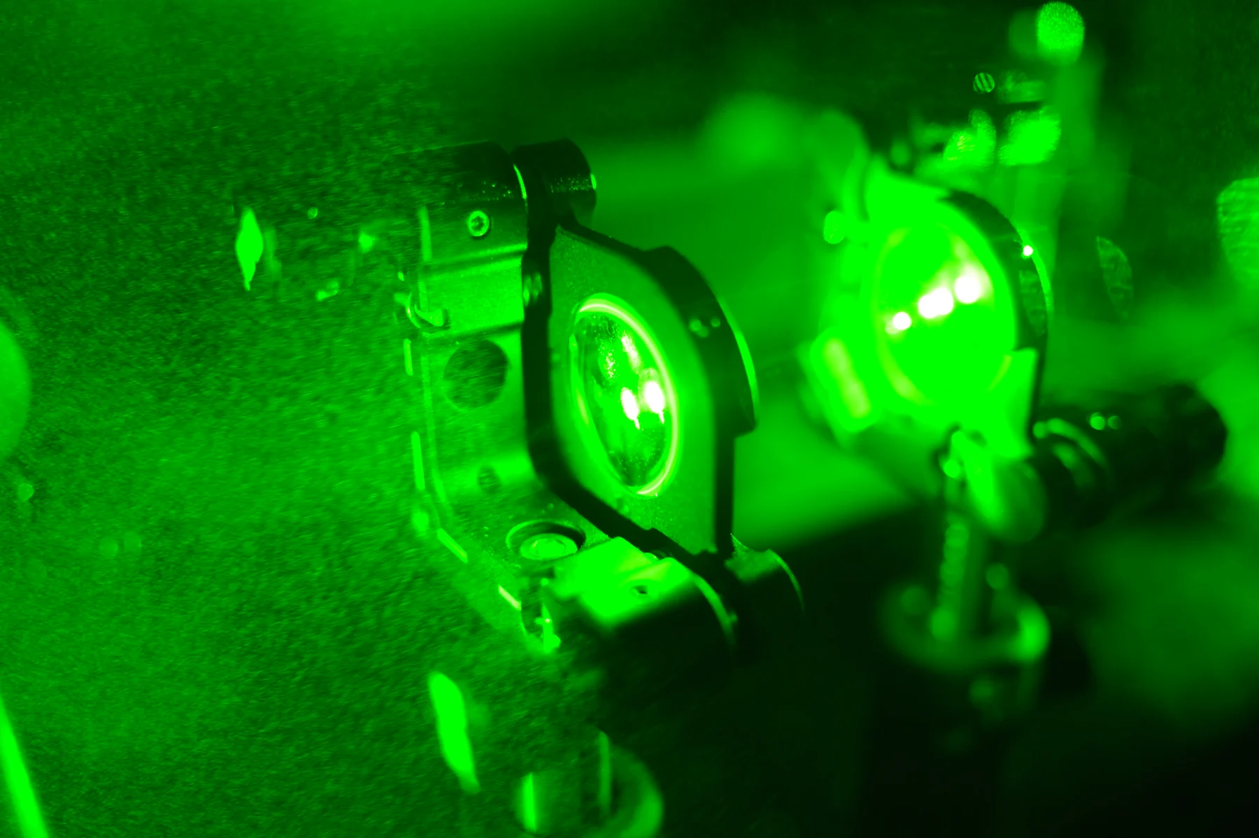
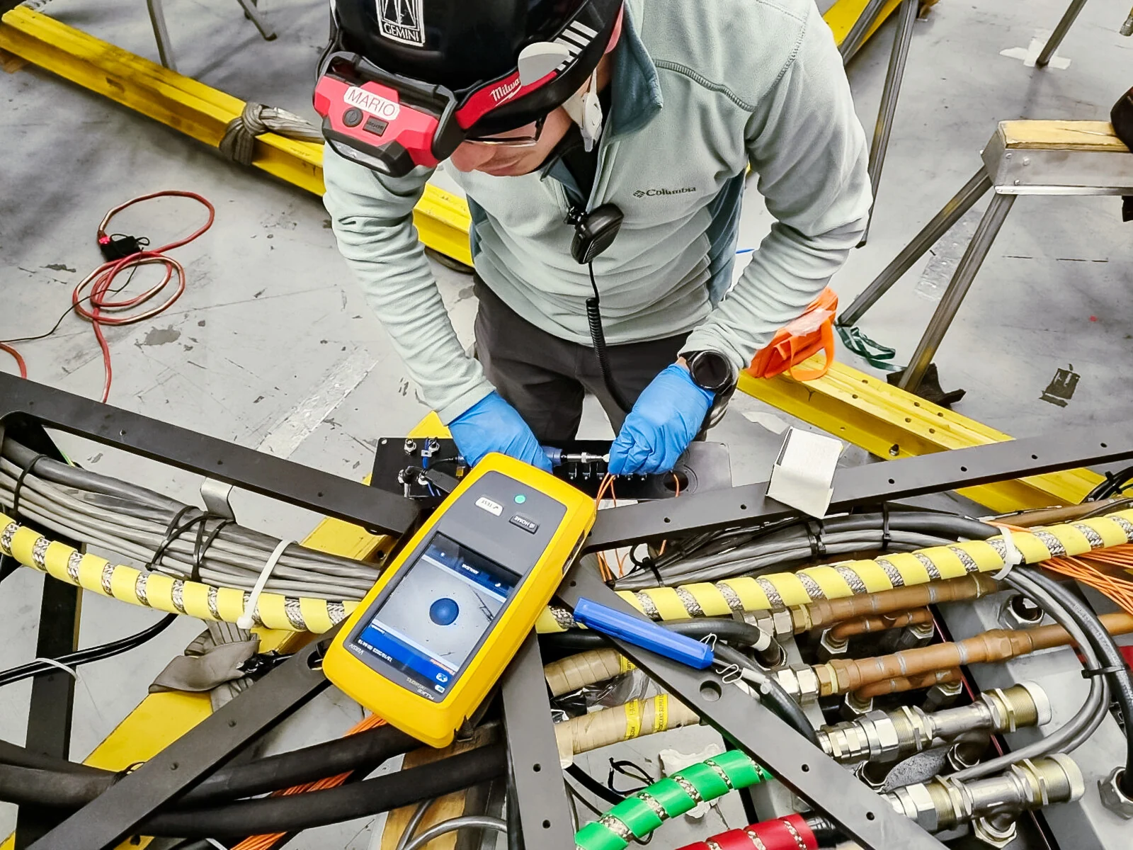
What must happen between shining light on tissue and seeing a useful medical image? Light has to reach a structure, interact with it, and return or generate a signal that a detector can capture. A lens or reconstruction method then turns that signal into information. Thinking through the whole path—from source to detector—shows why optical imaging can reveal some tissue properties in detail while struggling to see deep through the body.
The source sets the light in motion
An optical system begins with illumination or an existing source of light. The source may provide visible or infrared light, or deliver brief laser pulses. The choice matters because different wavelengths interact differently with biological materials. Light can be absorbed, scattered, reflected, or emitted, and those interactions carry information about the specimen [1].
In microscopy, light and lenses magnify small structures, allowing pathologists to examine tissue samples and researchers to study cells. In optical coherence tomography (OCT), infrared light is used to make subsurface images, including detailed views within the retina. The source does not create an image on its own; it supplies the energy that a tissue structure modifies in a measurable way [1].
Tissue changes the signal
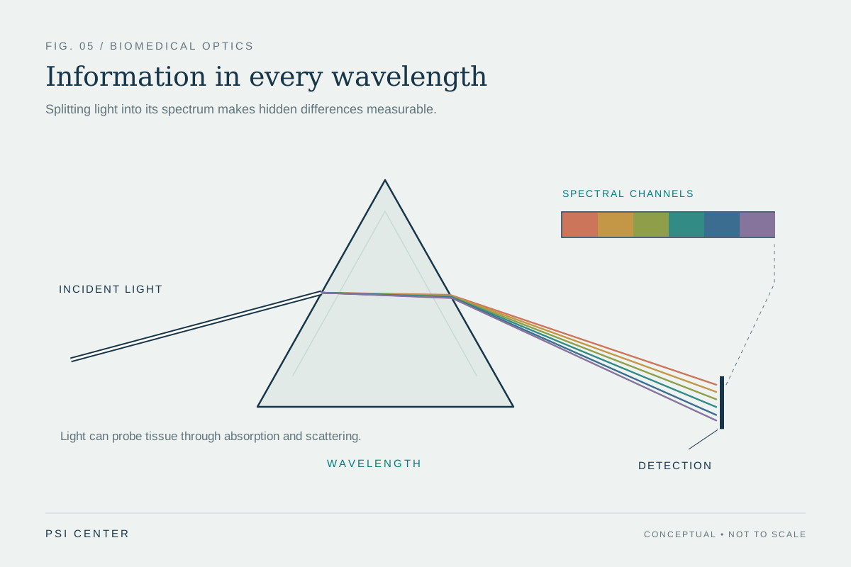
As light travels through tissue, molecules and structures alter its path. Absorption removes some wavelengths from the returning signal; scattering redirects light in many directions. Those changes can distinguish soft tissues or reveal properties such as blood oxygen saturation, but they also blur spatial information as light travels farther. This is one reason optical methods can be especially useful in soft tissue and for relatively shallow structures [1].
A useful contrast is ultrasound. Ultrasound forms images from sound echoes at tissue boundaries, while OCT uses reflected infrared light to image subsurface structure. Photoacoustic imaging couples the two: laser pulses are absorbed by tissue, producing ultrasound waves that a transducer detects. The optical input and acoustic detection together allow high-resolution views deeper than light-only methods can often provide [1].
Collection turns interaction into data
After interacting with tissue, light must reach a detector. In an endoscope, fiber optics and a miniature camera carry and collect light from internal cavities such as the stomach. In diffuse optical imaging, detectors collect near-infrared signals that have traveled through soft tissue; analysis can estimate properties such as total hemoglobin concentration and blood oxygenation [1].
The detector’s job is to convert incoming light into a measurable signal. It does not independently know whether the signal represents a vessel, a tissue boundary, or noise. Lenses, sensor placement, calibration, and later computation affect what can be reconstructed. The whole instrument therefore includes illumination, the tissue path, collection optics, sensors, and processing—not merely the lamp or laser [1] [2].
An image is an interpretation of measurements
Once collected, detector signals are processed into an image or map. A microscope magnifies a field of view; OCT produces cross-sectional information; photoacoustic methods reconstruct ultrasound signals generated by absorbed light. Each representation is shaped by what the system measured. For example, a map of oxygenation is not simply a photograph of a vessel: it is an estimate derived from light’s interaction with blood and tissue [1].
This full-system view also explains why optical imaging can be combined with other methods. MRI or ultrasound may contribute information that light alone cannot provide, while optical methods can measure tissue properties that other modalities do not. Repeated optical imaging can be useful because visible, ultraviolet, and infrared light are less hazardous than ionizing radiation, but the technique still must be matched to the question and tissue. No detector can recover information that never reaches it [1] [2].
Explore the physics: an explanatory diagram
Sources & further reading
- Optical Imaging NIBIB
- Sensors NIBIB
Prepared for the independent PSI Center reading library. About edition dates and source checking.
This explainer describes scientific principles. It does not assess individual treatment choices.