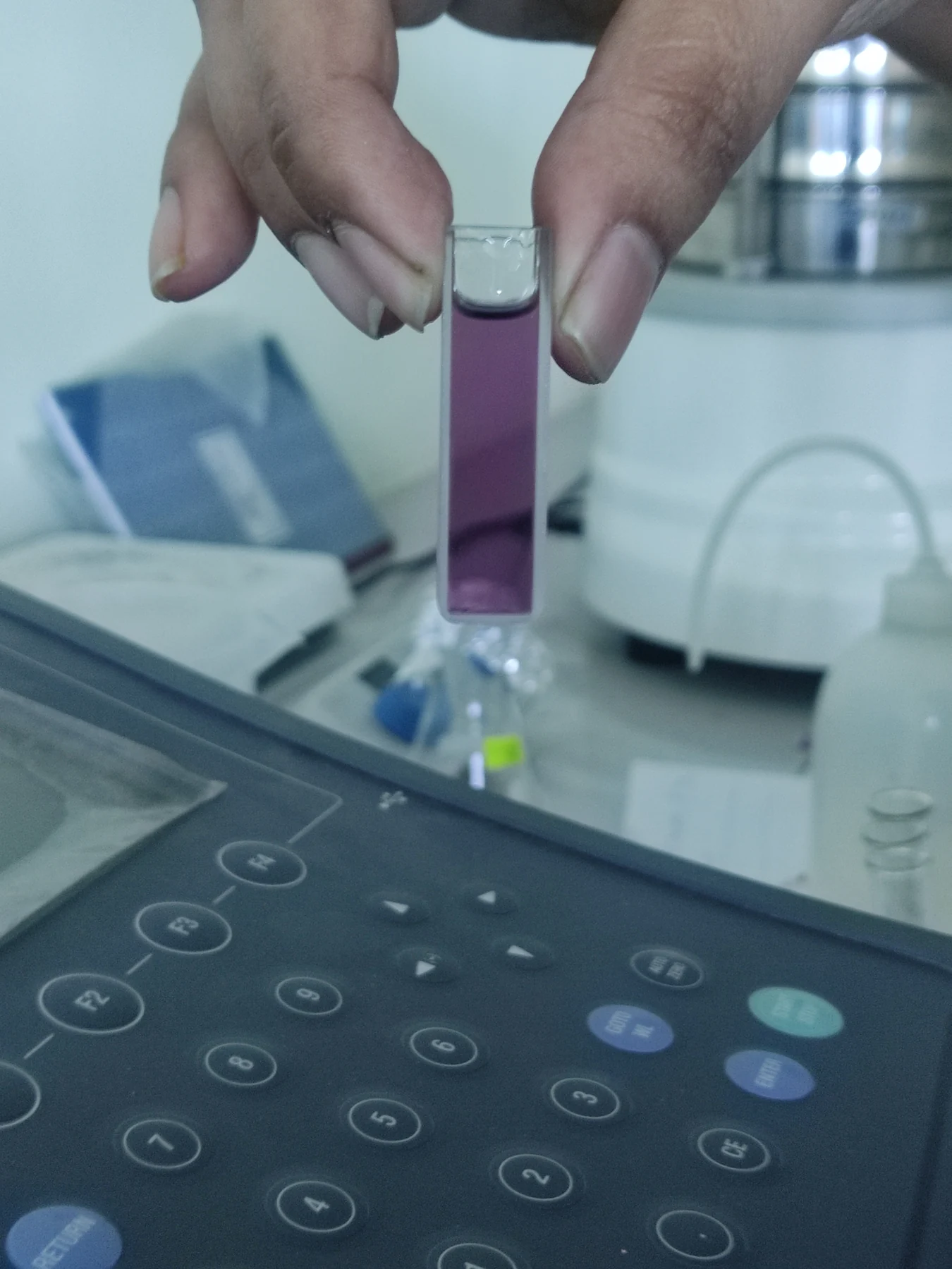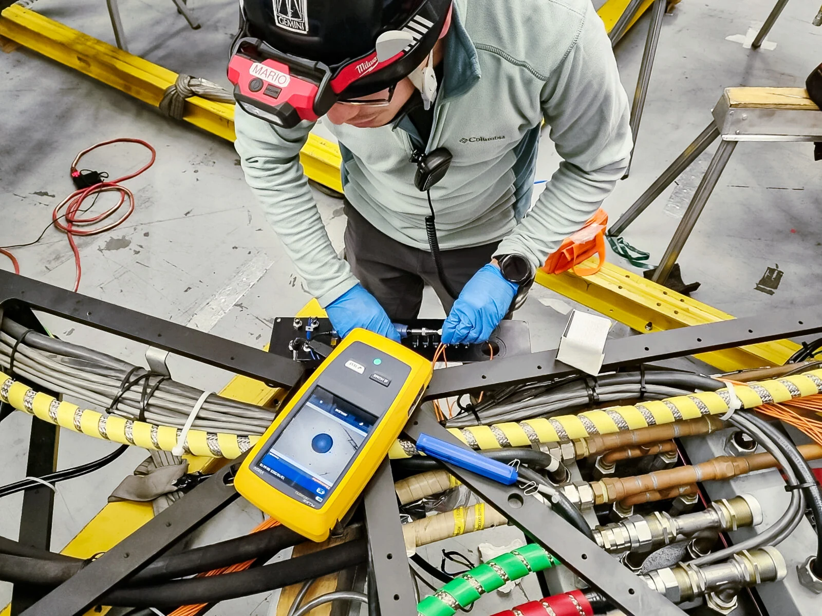
If a phone camera can show a pulse in a fingertip video, can it replace a medical sensor? A camera is a sensor too, but it measures light arriving at its pixels. Turning that signal into a claim about oxygen, glucose or heart function requires additional steps: a model of the body, calibration and testing against an appropriate reference. The distinction is between seeing a changing pattern and measuring a health variable reliably enough for a defined use.
Every sensor has a specific input
A sensor detects and responds to an input from the physical environment. Some measure temperature, pressure, flow or chemical properties; optical sensors detect light. In health care, different devices are designed to detect particular biological, chemical or physical processes and report data to a person or clinician. A thermometer, for example, translates a physical change caused by heat into a value associated with temperature. [1]
A camera’s pixels register light intensity and color over time. In a fingertip video, changing blood volume can alter the amount of light reflected, producing a periodic pattern. But that observation alone is not an oxygen measurement. A pulse oximeter is designed to use changes in absorption of particular types of light to estimate heart rate and blood oxygen. Its optical pathway and interpretation target a defined signal. Similar-looking sensors can therefore answer different questions. [1]
From signal to health claim
The inference chain matters. First, light interacts with tissue; then a detector records a signal; software separates a pattern from motion, lighting changes and noise; finally, an algorithm maps that pattern to a physiological quantity. Each step can introduce error. A hand moving, ambient light shifting, skin properties varying, or the camera changing exposure may alter pixels without a corresponding change in the target variable. A model must distinguish these effects from the biological signal it is meant to estimate.
A mental experiment makes the issue clear: film the same finger under a lamp, then in daylight, while keeping the pulse similar. The raw colors may differ because illumination and camera settings differ. A robust measurement method needs ways to control or account for such variation. It also needs comparison with a trusted reference across relevant conditions. Without that validation, a plausible number displayed on a screen is still an estimate whose accuracy and limits are unknown.
Validation belongs to the intended use
Medical devices can monitor, modulate or supplement bodily functions, and they range from implanted pacemakers to external wearable sensors. Some wearables track heart rate or sleep patterns; continuous glucose monitors use a sensor that detects glucose in fluid between cells. These examples show that a device’s role depends on what its sensor is built to detect and how the result is used. They do not imply that every consumer wearable diagnoses disease. [2] [1]
Validation asks whether the device performs as intended in the context where people will use it. A method tested in controlled lighting may behave differently during exercise, in a dim room or across different camera hardware. A device intended to flag a possible change has a different task from one intended to establish a diagnosis. Evidence must match the claim, population and setting; an algorithm that identifies a pattern in a dataset is not automatically a clinically validated instrument.
Why the instrument’s design matters
Some devices connect measurement to action. NIBIB describes continuous glucose monitoring paired with an insulin pump that can adjust insulin delivery when glucose falls too low. This illustrates a system with a defined sensor, a linked signal pathway and a specific function. The safety and usefulness of such a system depend on the parts working together; a camera’s ability to detect color variation does not by itself recreate that system. [2]
Cameras can support research and may contribute to useful measurement tools when their signals are carefully interpreted and tested. The point is not that optical methods are inherently weak: pulse oximeters themselves rely on light. The distinction is whether the complete measurement system has a clear target, reliable signal processing and evidence for its intended use. A camera can collect data; a validated medical sensor is a tested instrument whose readings have been shown to mean what the device claims they mean.
Sources & further reading
- Sensors NIBIB
- Medical Device Technologies NIBIB
Prepared for the independent PSI Center reading library. About edition dates and source checking.
This explainer describes scientific principles. It does not assess individual treatment choices.