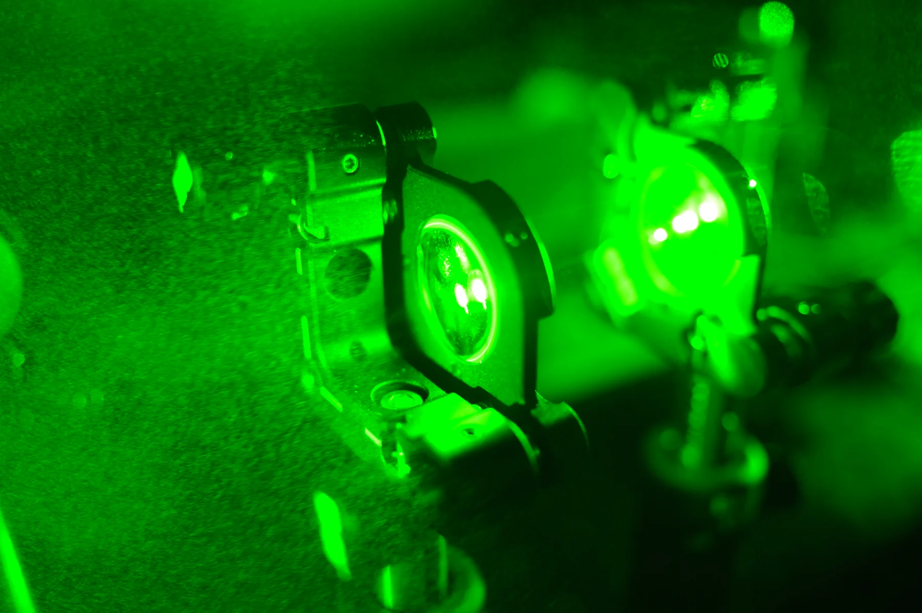
If a fluorescent dye glows, why doesn’t a detector record only the dye? Light entering biological tissue can scatter, reflect, or be absorbed, and some molecules emit light of their own. A detector collects photons from the whole optical path, not just from the intended label. The resulting background is part of the measurement: it can obscure a weak signal, but its sources can also reveal what the sample is doing.
Fluorescence is a change in light
A fluorescent molecule absorbs light at one range of wavelengths and then emits some of that energy as light at a different wavelength. In an imaging setup, illumination excites the label, and filters help a detector collect the emitted light while rejecting much of the illumination. The signal is useful because it can mark a molecule, cell, or tissue feature.
But the label is only one possible emitter. Biological samples contain many molecules, and some produce autofluorescence: light emission without an added fluorescent label. The detector cannot inherently know which photons came from the chosen dye and which came from the sample itself. Optical imaging relies on light’s absorption, scattering, reflection, and emission in combination [1].
Light takes indirect paths through tissue
A photon entering tissue may scatter, changing direction after interactions with microscopic structures. It may be absorbed, or reflected back toward the surface. Scattering can blur the origin of detected light because photons reaching the detector may have traveled along different paths from those expected in a simple straight-line picture [1].
Imagine shining a narrow flashlight beam into fog. Some light reaches the target, but droplets also scatter light toward your eyes from many locations. Tissue is more complex than fog, yet the analogy captures why a bright illumination source can produce a diffuse background around a desired feature.
Sensors register photons, then infer meaning
An optical sensor converts incoming light into an electrical signal. It counts or measures photons within selected wavelength bands, but the raw reading reflects all light that passes through the optical system. Background may come from tissue autofluorescence, scattered excitation light, reflections, or detector and instrument effects. Separating these contributions requires design choices such as filters, reference measurements, and image processing.
Biomedical sensors can also report signals from fluorescent probes whose brightness changes with a chemical condition. NIBIB describes research nanosensors that emit fluorescence in response to drug concentration, with changes detected through the skin in animal experiments [2]. That example illustrates a key distinction: the sensor’s emitted signal can encode a quantity, while tissue and optics still shape how much of that signal reaches a detector.
Background can limit contrast and carry clues
The ratio between a feature’s signal and surrounding background affects whether the feature is visible. If both rise together, simply increasing illumination may not clarify the image; it can also increase unwanted light or potentially affect the sample. Researchers instead choose wavelengths and detection geometry suited to the tissue, and may compare measurements made with and without a fluorescent label.
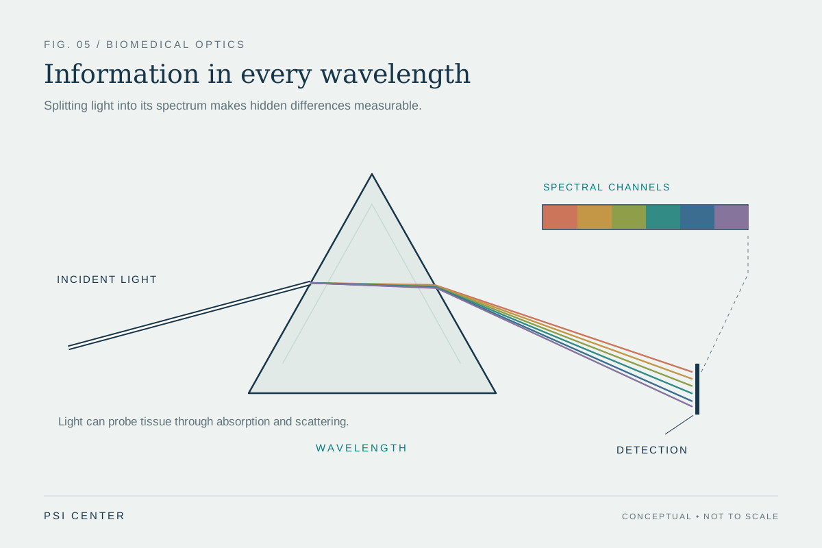
Background is not always meaningless noise. Optical methods exploit differences in how tissues absorb and scatter light to measure properties such as blood oxygenation, and some techniques combine light with ultrasound to image deeper structures [1]. Still, a bright optical signal alone does not establish a diagnosis. It is a measurement shaped by the probe, tissue, and detector, and its biological meaning depends on validation and context.
Explore the physics: an explanatory diagram
Sources & further reading
- Optical Imaging NIBIB
- Sensors NIBIB
Prepared for the independent PSI Center reading library. About edition dates and source checking.
This explainer describes scientific principles. It does not assess individual treatment choices.