How can a flash of light help form an image deep inside tissue, where light itself scatters? Photoacoustic imaging uses a short laser pulse to deliver energy to tissue. Where molecules absorb that light, they warm slightly and expand for an instant. That small expansion launches an ultrasound wave, which travels outward to a detector. The method begins with light but reads out sound, combining optical sensitivity with the ability of acoustic waves to carry information from within the body.
Absorption makes a local heat pulse
Light does not interact with all tissues in the same way. Some molecules absorb particular wavelengths more strongly than others, while tissue can also scatter, reflect, or transmit light [1]. In photoacoustic imaging, a brief laser pulse is chosen so that absorbing structures take up some of its energy. Blood is often a useful target because hemoglobin absorbs light, and its oxygenation state affects optical absorption.
The absorbed energy becomes heat, but the pulse is brief and the intended temperature rise is small. The heated region expands slightly; when it cools, it contracts. This rapid expansion creates a pressure disturbance that propagates as sound. The key chain is light absorption, transient heating, expansion, and an acoustic wave—not a camera directly seeing a deep structure through tissue.
A transducer listens for the wave
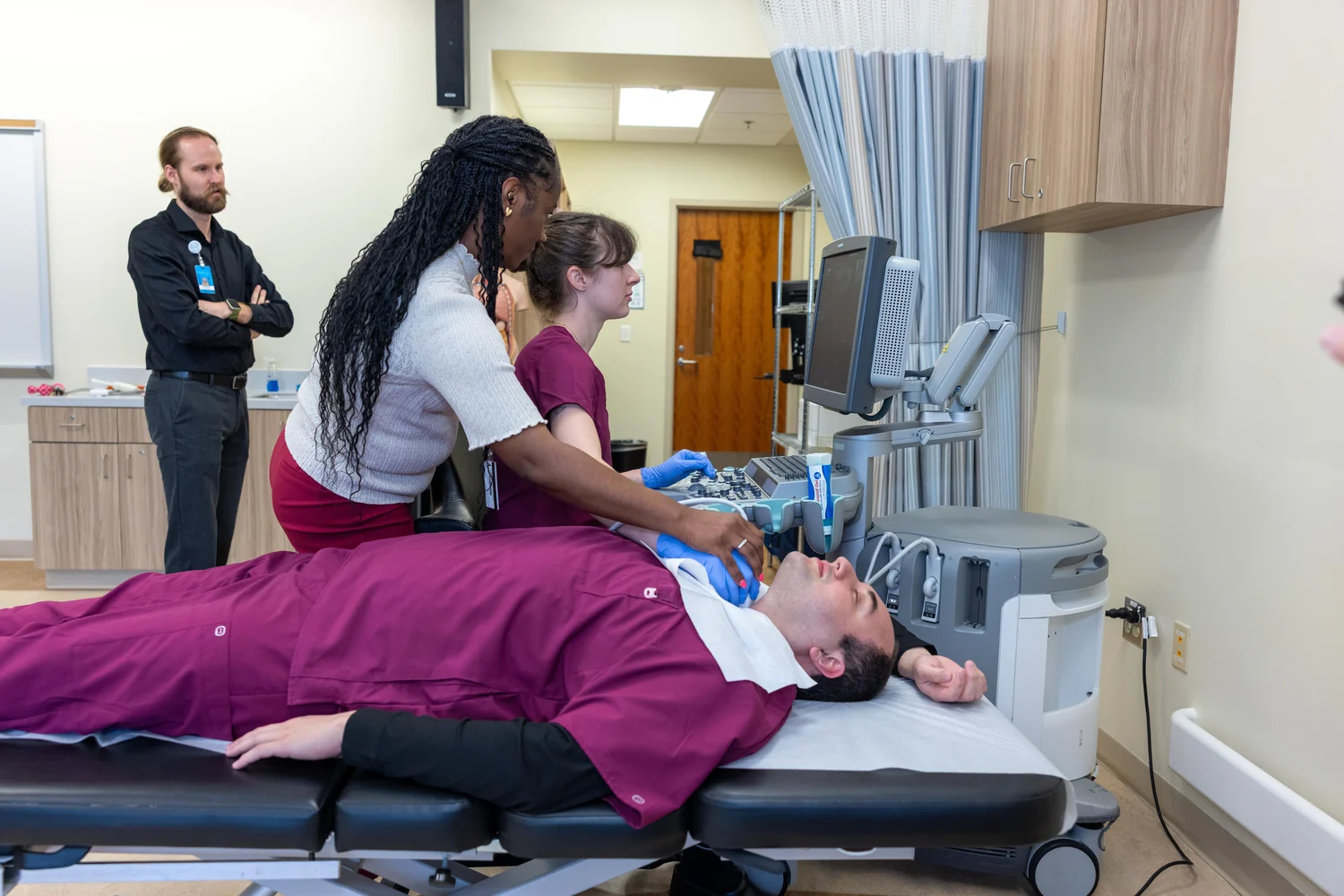
An ultrasound transducer detects returning pressure waves and converts them into electrical signals. In conventional ultrasound, the transducer sends sound into the body and records echoes reflected at boundaries between tissues. Photoacoustic imaging reverses the source of the sound: tissue generates the wave after absorbing light, and the transducer listens [2].
The arrival times and strengths of the waves help estimate where absorbing structures are located. A sound wave that arrives sooner generally came from a nearer source, while signals from different positions can be combined to form an image. Since tissue scatters light more strongly than it scatters ultrasound in many relevant settings, acoustic detection can preserve spatial information from sources that would be difficult to image optically at depth.
What makes the hybrid useful
Optical methods can be sensitive to molecular composition, while ultrasound can locate structures through acoustic reflections and timing. Photoacoustic imaging joins these strengths. Depending on wavelength and method, researchers can map blood vessels, study blood oxygenation, or examine other light-absorbing features. NIBIB describes potential uses that include monitoring vessel growth in tumors, detecting skin melanomas, and tracking oxygenation in tissue [1].
A mental experiment helps distinguish the signal from a photograph. Imagine shining a brief, controlled flash on a dark patch hidden beneath translucent layers. The patch absorbs more energy, expands minutely, and emits a sound pulse. The detector does not record the patch’s visible color; it infers the absorbing source from the wave that travels back. Varying the light wavelength can change which molecules absorb most, providing another kind of contrast.
Promise and limits in practice
A method’s ability to create an image does not establish that it improves clinical outcomes. The source describes photoacoustic imaging as an imaging approach and reports research systems that have visualized blood flow in human hands and feet, with hopes for other applications [1]. Those research demonstrations do not mean every proposed use is established routine care.
Image quality depends on how much light reaches a target, how strongly relevant tissue absorbs it, how sound travels back, and how the system reconstructs the measurements. Tissue depth and optical scattering constrain the light delivery; acoustic boundaries and noise affect detection. Photoacoustic imaging is therefore a useful example of a broader strategy in medical physics: convert one form of energy into another that can carry a measurable signal, then interpret that signal carefully rather than treating it as a direct view of disease.
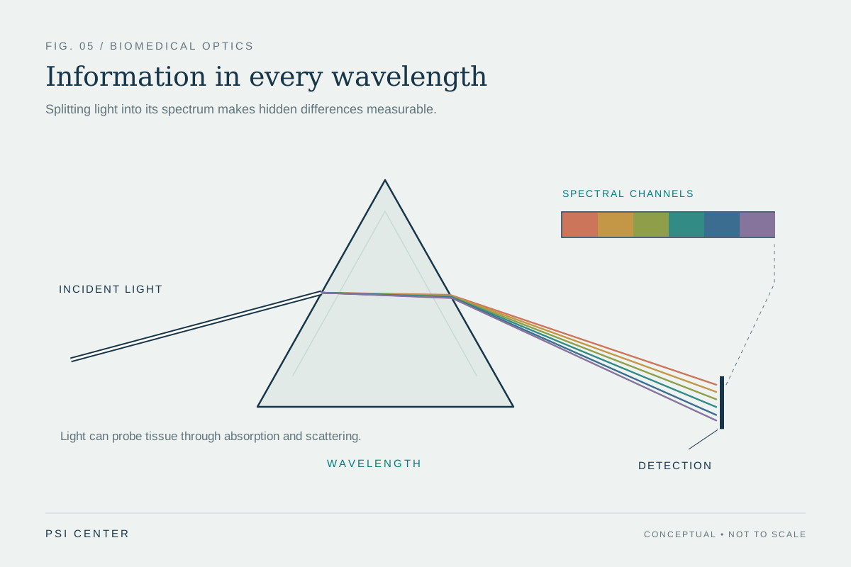
Explore the physics: an explanatory diagram
Sources & further reading
- Optical Imaging NIBIB
- Ultrasound NIBIB
Prepared for the independent PSI Center reading library. About edition dates and source checking.
This explainer describes scientific principles. It does not assess individual treatment choices.