How can a beam of light reveal layers inside the eye without cutting into them? Optical coherence tomography, or OCT, measures light reflected from tissue at different depths and assembles those returns into a cross-sectional image. The method resembles ultrasound in its use of echoes, but the signal is light rather than sound. Its success depends on a useful balance: light can resolve fine structure, yet tissue also scatters and absorbs it.
Light returns carry depth clues
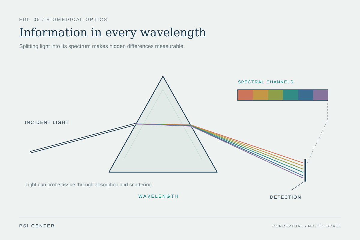
Light changes when it meets biological material: it can be absorbed, scattered, reflected, or emitted. Optical imaging uses those interactions to visualize tissues and structures. OCT is one such method, using infrared light to obtain subsurface images of tissue [1].
A useful mental picture is a stack of faint reflections. Light returning from near the surface arrives differently from light that has traveled farther into tissue and back. OCT analyzes these depth-dependent returns and constructs a cross-section. The analogy to ultrasound helps explain the geometry—both methods infer internal structure from returning signals—but their physical waves differ: ultrasound sends sound; OCT sends light [1].
Why the retina is a natural target
The retina is a light-sensitive layer at the back of the eye. In normal vision, light passes through the cornea and lens and reaches the retina, where photoreceptors convert it into electrical signals that travel along the optic nerve to the brain [2]. OCT uses reflected infrared light to image retinal structure in detail, giving ophthalmologists a view of layers that cannot be seen directly from the eye’s front surface [1].
The eye offers a particularly useful setting for optical imaging because light can enter and return through transparent structures before reaching the retina. In other tissues, scattering can blur or weaken signals with depth. OCT’s strength is therefore not that light penetrates everywhere, but that it can reveal fine subsurface organization where the optical path is workable.
A cross-section is a sampled view
An OCT image is not the tissue itself; it is a reconstruction from reflected signals. Like any imaging method, it turns physical measurements into a visual representation. Changes in reflection can indicate boundaries and structure, but interpreting what those structures mean requires medical context and expertise. The measurement does not independently diagnose every condition.
OCT is also used by cardiologists to help diagnose coronary artery disease, showing that the technique is not limited to eye care [1]. Yet the same instrument principle does not make every organ equally accessible. Light’s interaction with the particular tissue, the distance to the target, and the access route shape what can be captured.
Optical images alongside other evidence
Different imaging methods answer different questions. OCT supplies detailed structural views near a surface; other optical approaches can estimate tissue properties such as blood oxygenation or combine light with ultrasound to image deeper regions [1]. In research and care, optical imaging can also be combined with methods such as MRI to add complementary information [1].
The distinction is useful when reading an image: seeing a layer or boundary is not the same as measuring how an organ functions, and neither alone proves what treatment will work. OCT is a way to make otherwise hidden structure visible through reflected light. Its value comes from matching that physical measurement to a specific clinical question, such as examining retinal organization.
Explore the physics: an explanatory diagram
Sources & further reading
- Optical Imaging NIBIB
- How the Eyes Work National Eye Institute
Prepared for the independent PSI Center reading library. About edition dates and source checking.
This explainer describes scientific principles. It does not assess individual treatment choices.