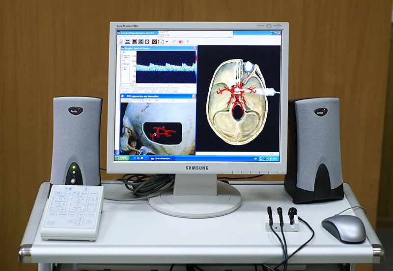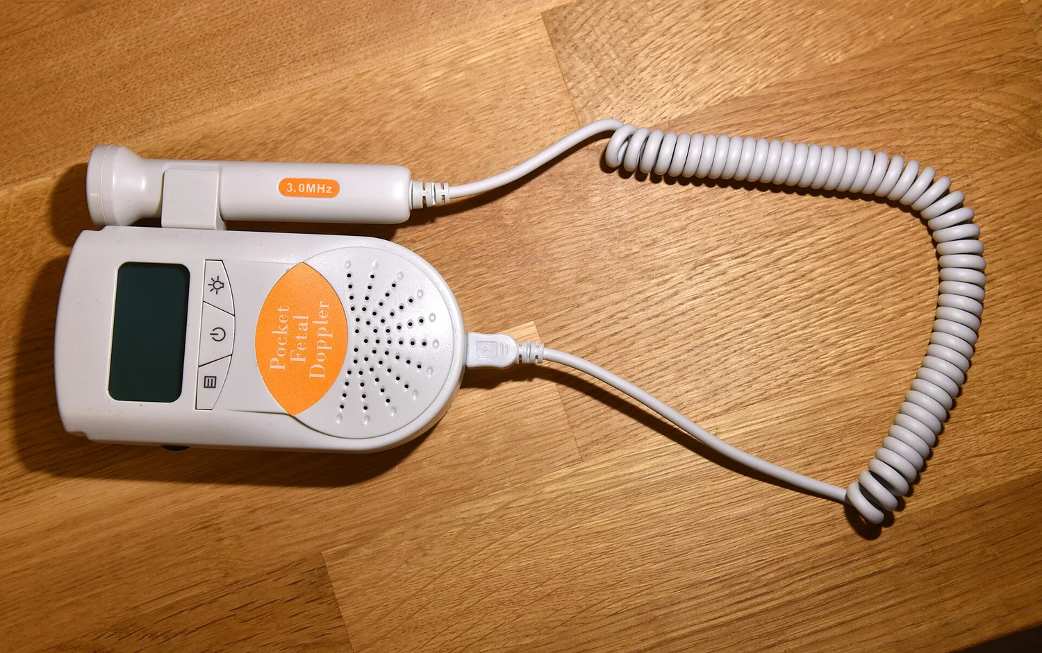
If two sound pulses have different amplitudes, do they necessarily carry different intensity? The quantities are related, but they describe different things. Amplitude measures the size of a changing signal, while intensity concerns energy flow through an area. Ultrasound imaging relies on echoes whose amplitudes help form an image, yet the energy delivered to tissue is a separate physical consideration.
A wave has several measurable features
Picture a sound wave as pressure fluctuations traveling through tissue. Its amplitude describes the size of those fluctuations relative to the surrounding pressure. A larger amplitude means a stronger oscillation in the measured signal. Frequency, by contrast, counts how many oscillations occur per second; it is not another name for amplitude.
Intensity describes the rate of energy flow per unit area. For a simple traveling wave under fixed conditions, intensity rises with the square of amplitude: doubling amplitude corresponds to four times the intensity. This relation is useful as a mental model, but actual ultrasound fields and tissue interactions can be more complex than a single uniform wave.
Echo amplitude helps build an image
In diagnostic ultrasound, a transducer sends high-frequency sound into the body and receives echoes reflected from tissue boundaries. The scanner uses the echo’s travel time to estimate depth and the signal strength, including amplitude, to help produce an image [2]. A stronger returning echo can appear brighter in the display, though image processing and settings also shape the final appearance.
This is different from saying that a bright pixel directly reports the intensity that entered tissue. The detected echo has traveled through tissue, reflected, and returned; it is a received signal. The original transmitted field and energy deposition at a location are related questions, but they are not the same measurement.
From sound energy to tissue effects
Ultrasound is non-ionizing: it uses sound waves rather than the ionizing radiation used in x-ray imaging. Diagnostic ultrasound is generally considered safe when used prudently by trained providers, but ultrasound energy can produce biological effects, including slight heating and mechanical effects such as cavitation under some conditions [2]. These effects depend on the field and exposure, not simply on how bright an image looks.
NIBIB distinguishes diagnostic ultrasound, which creates images, from therapeutic ultrasound, which can use high-intensity beams to heat, move, or destroy tissue [1]. The distinction makes physical sense: the same broad kind of wave can be used at different operating conditions and for different purposes. A diagnostic echo display is not a map of therapeutic energy.
Why keeping the terms separate matters
Suppose a scanner receives a stronger echo from one boundary than another. That tells the system something about the reflected signal, helping it represent anatomy. It does not, by itself, mean that the tissue absorbed more energy or experienced a greater effect. Reflection, transmission, attenuation, beam shape, and duration all influence what happens to energy as sound moves through the body.
The FDA describes ultrasound’s ability to heat tissue slightly and produce pressure oscillations with mechanical effects, and recommends minimizing exposure while maintaining diagnostic quality [2]. Amplitude and intensity give different views of the wave: one describes signal size, the other energy flow. Treating them as interchangeable obscures both how an image is formed and how ultrasound interacts with tissue.
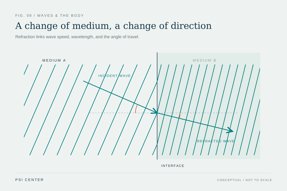
Explore the physics: an explanatory diagram
Sources & further reading
- Ultrasound NIBIB
- Ultrasound Imaging FDA
Prepared for the independent PSI Center reading library. About edition dates and source checking.
This explainer describes scientific principles. It does not assess individual treatment choices.