Why does a signal sent into the body come back weaker than it went in? Energy can be absorbed, scattered, or redirected before it reaches a detector. The result is attenuation: a gradual loss of signal strength with distance. It is not a single physical process, and comparing light with ultrasound shows why tissue depth matters so much to medical imaging.
Energy takes several routes out
Imagine a flashlight shining through a glass of milk. The beam becomes dim because some light is absorbed: its energy is transferred to the material. Other photons are scattered, changing direction after interactions with structures inside the liquid. A detector placed straight ahead sees less light, even though some of that light still exists elsewhere. Biological tissue can absorb, scatter, reflect, or emit light, and optical imaging relies on these interactions to reveal features at scales from molecules to organs [1].
Absorption and scattering have different consequences. Absorbed energy may contribute to heating or drive a chemical process; scattered energy may carry information but no longer travel along the path the detector expects. Reflection at a boundary can also send a signal back toward its source. When a measurement counts only energy arriving in a particular direction, any process that diverts it appears as signal loss.
Sound echoes are not lossless
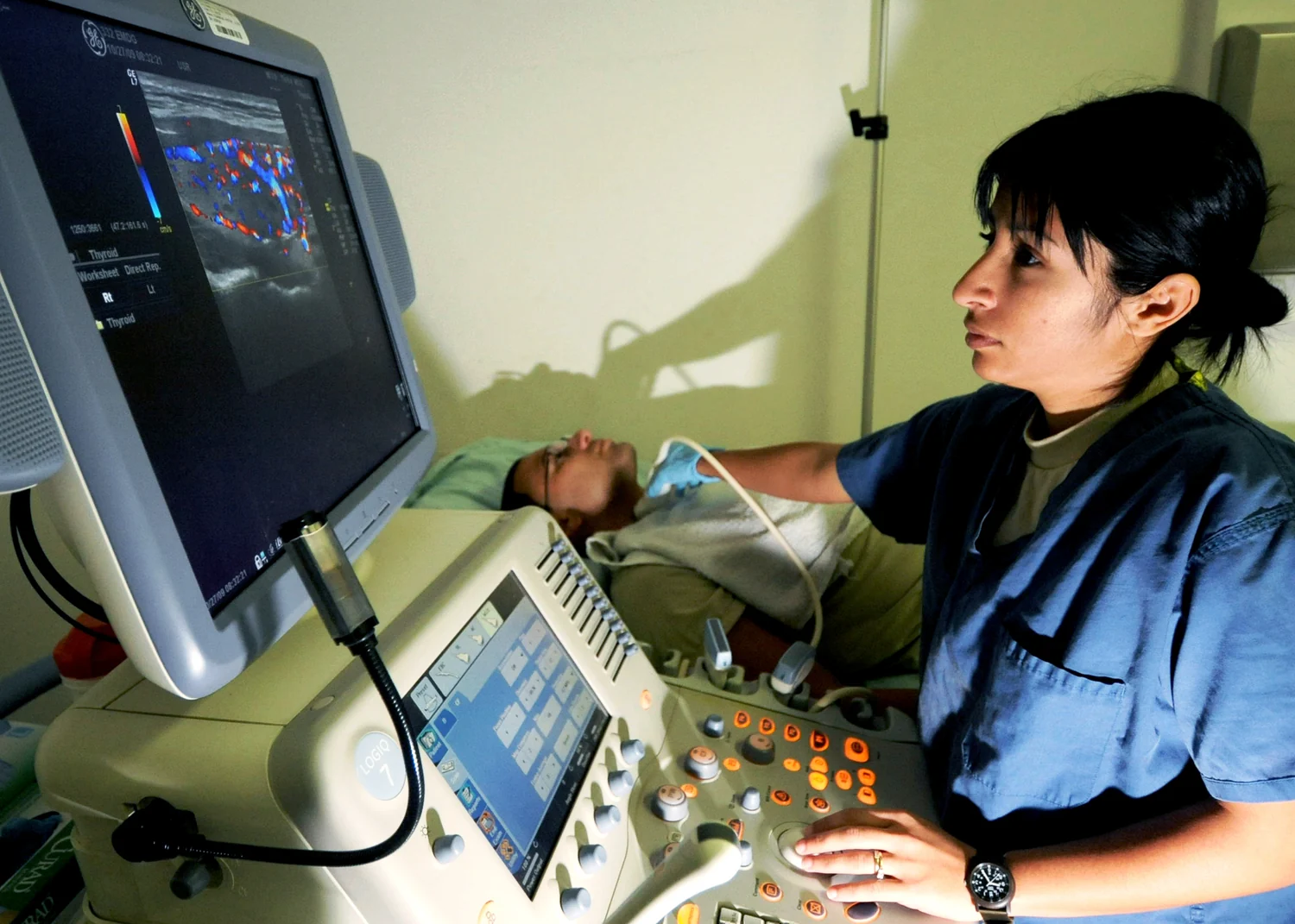
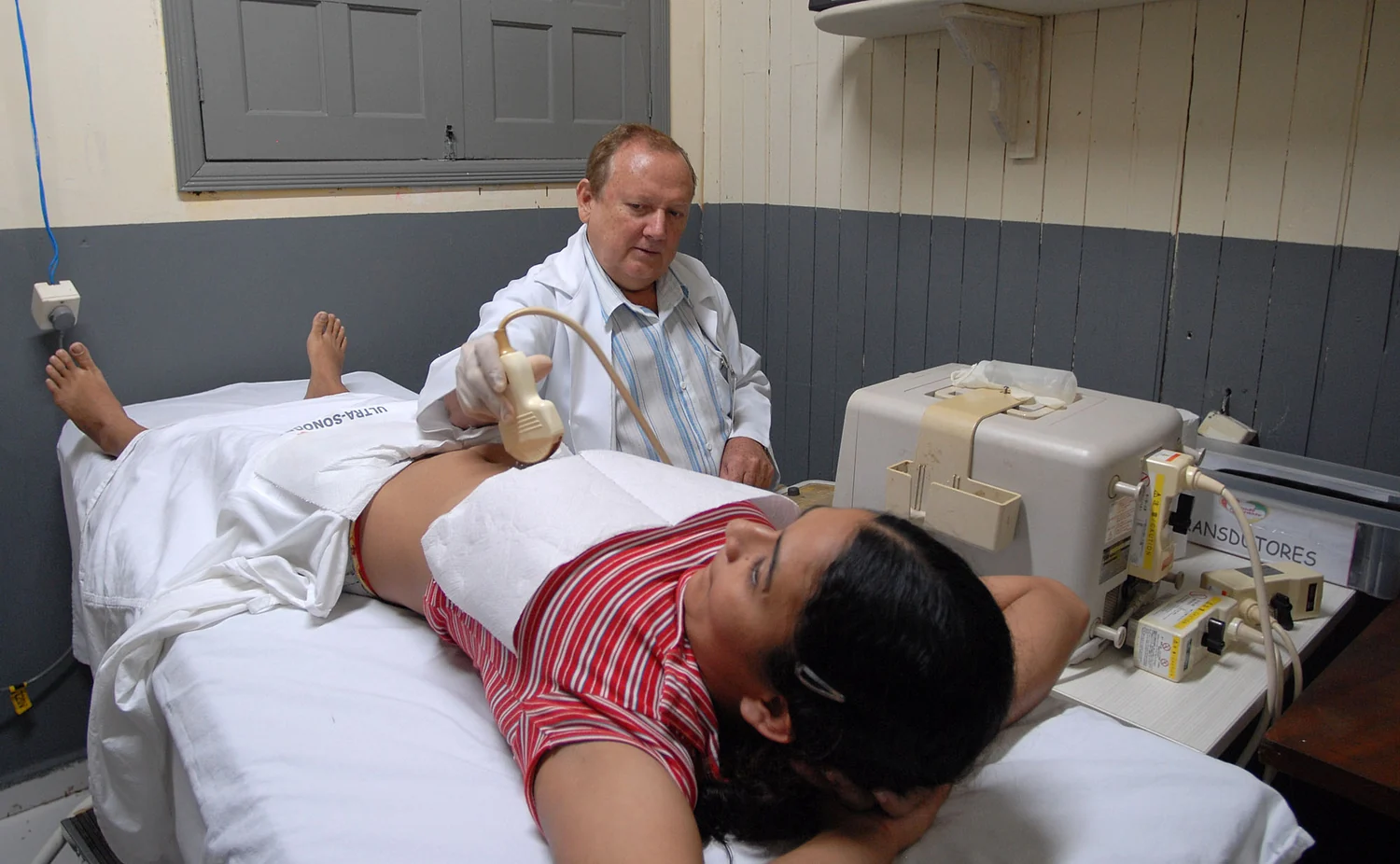
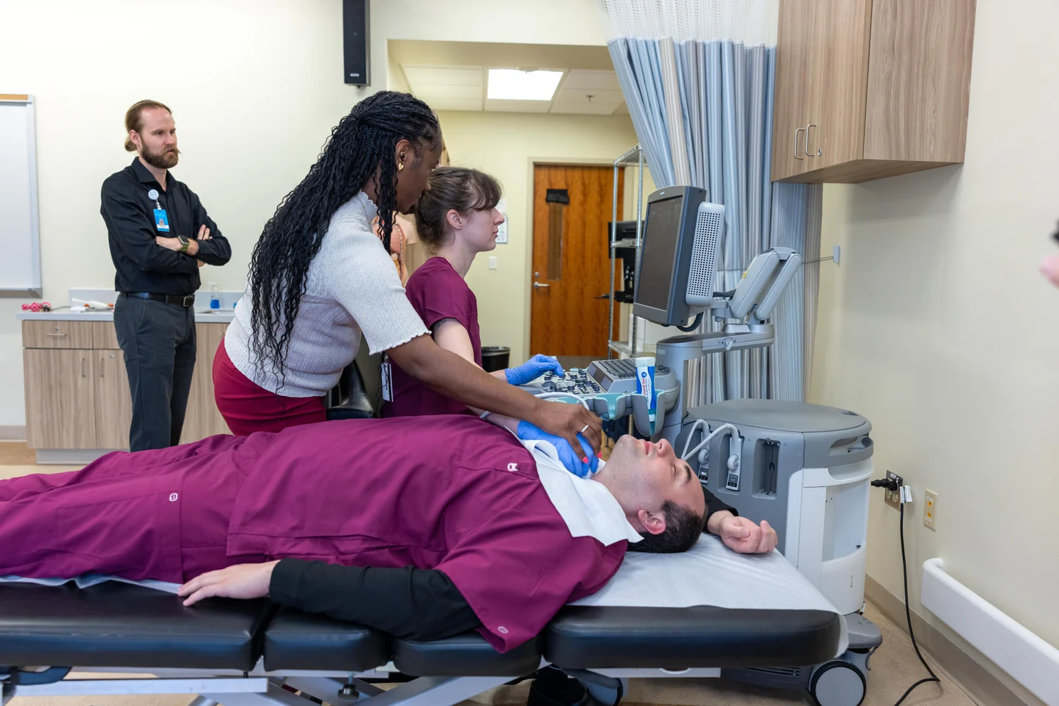
Ultrasound provides a useful comparison. A transducer sends high-frequency sound into the body and listens for echoes from boundaries between tissues. The scanner estimates a boundary's depth from the time an echo takes to return. But sound can weaken as it travels, and some energy is reflected away or absorbed rather than returning to the probe. A weak echo from deep tissue therefore does not automatically mean the structure itself is faint or unimportant [2].
Consider two echoes from similar boundaries, one near the skin and one much deeper. The deeper echo has traveled farther on its outward and return journeys, so it has had more opportunity to lose energy. The scanner must distinguish that distance-related decline from real differences among tissues. In practice, the image is an interpretation of measured echoes, not a direct photograph of every structure.
Depth changes the optical picture
Light also faces a longer and more complicated journey at greater depth. Near-infrared light can be used in diffuse optical imaging to measure properties such as hemoglobin concentration and blood oxygen saturation in soft tissue [1]. The light that reaches a detector after passing through tissue has been filtered by absorption and scattered along many paths. Its remaining strength, timing, or distribution can help infer tissue properties, but the inference depends on how light behaved on the way.
This is one reason optical techniques suit some questions better than others. Optical coherence tomography uses infrared light to form subsurface images, including detailed views of the retina, while diffuse optical methods can measure soft-tissue properties. Photoacoustic imaging uses a different sequence: tissue absorbs laser pulses and generates ultrasound waves, which a transducer detects. The detected sound carries information about where optical energy was absorbed [1].
A measurement is a tradeoff
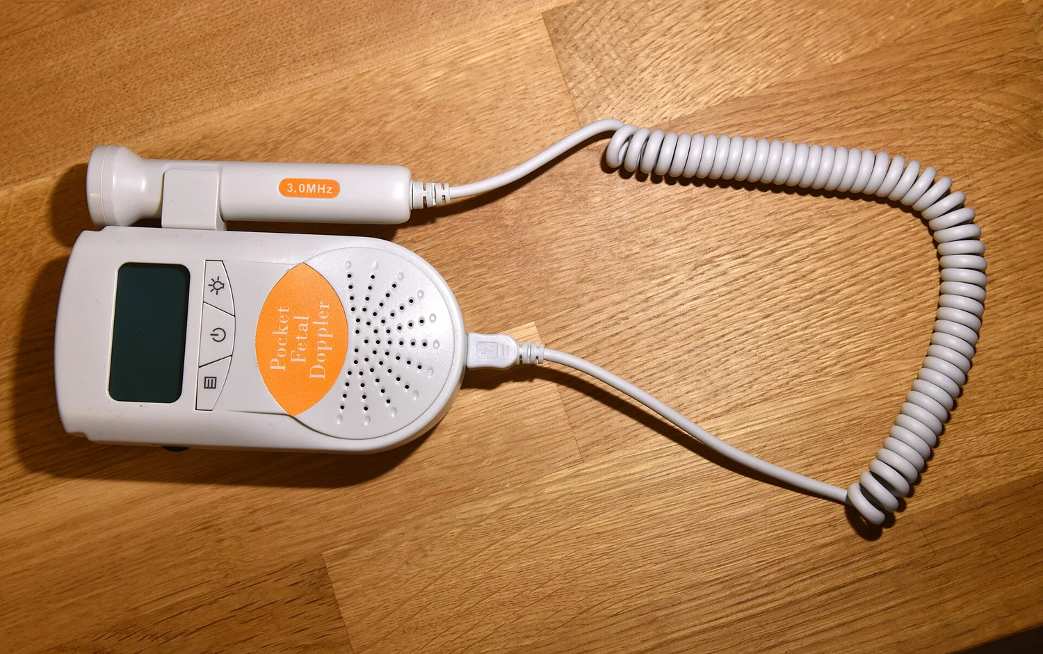
Frequency and wavelength matter because they change how signals interact with tissue. In ultrasound, the probe's operating frequency affects the signal and the echoes it can collect; the image also depends on the path through the body and the boundaries encountered [2]. Optical methods likewise use different parts of the light spectrum for different purposes. There is no universally best signal: an imaging method is chosen partly for the depth and tissue feature it needs to resolve.
A helpful mental experiment is to picture a row of listeners waiting for a single clap. Those closest hear it most clearly; farther listeners hear a quieter sound, and walls or other obstacles redirect some of it. Medical detectors face a more complex version of this problem, with energy entering and leaving tissue in many directions. Attenuation is both a limit and a clue: the missing signal constrains what can be seen, while the pattern of loss can itself reveal how the tissue is structured.
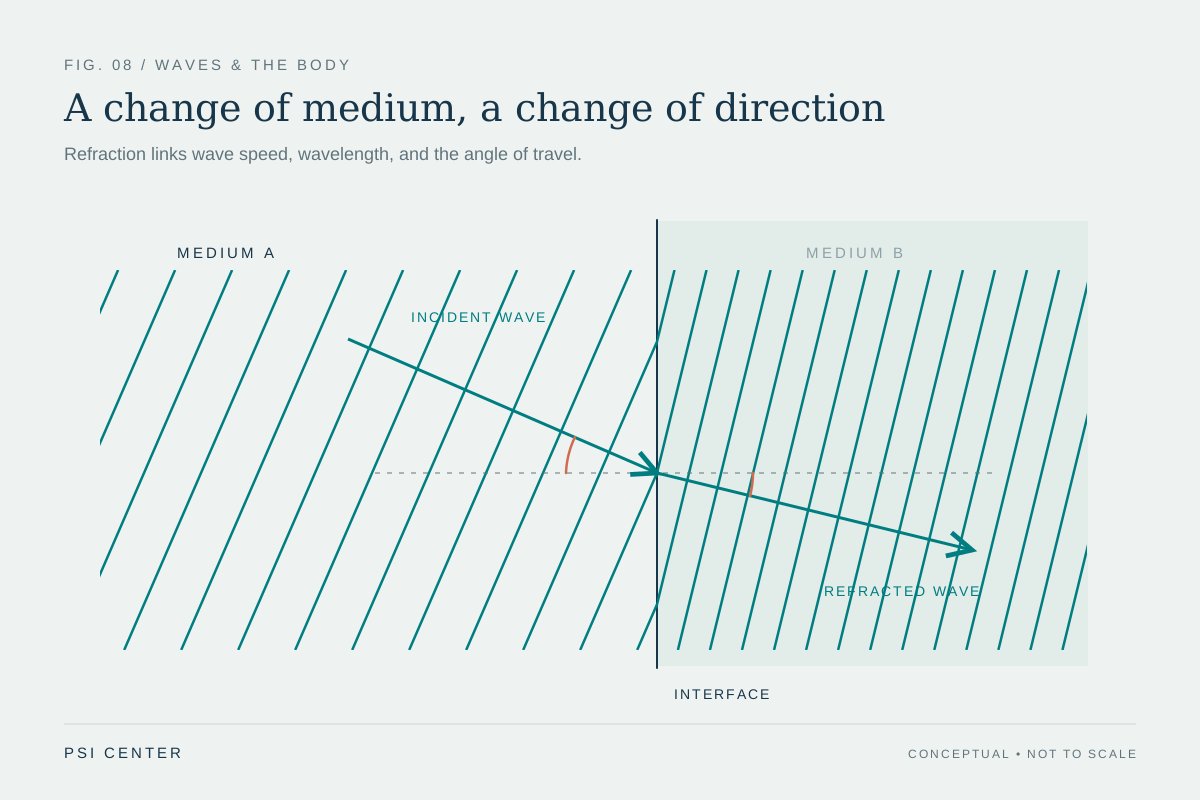
Explore the physics: an explanatory diagram
Sources & further reading
- Optical Imaging NIBIB
- Ultrasound NIBIB
Prepared for the independent PSI Center reading library. About edition dates and source checking.
This explainer describes scientific principles. It does not assess individual treatment choices.