What does it mean for a medical signal to have a high frequency? It means that its cycles repeat rapidly, not necessarily that the wave travels faster. Wavelength is the distance covered during one cycle. The two quantities are linked by the wave’s speed: if speed stays fixed, more cycles packed into each second mean less distance available for each cycle. That simple relationship helps make sense of both ultrasound images and the much wider electromagnetic spectrum.
Count the cycles, then measure their spacing
Frequency is the number of repeated cycles per second, measured in hertz (Hz). Wavelength is the distance from one point in a wave pattern to the next matching point, such as crest to crest. For a wave moving at a steady speed, frequency multiplied by wavelength equals speed. A swimmer making more strokes per second in a pool does not make the pool longer; likewise, if wave speed is fixed, a higher frequency corresponds to a shorter wavelength.
Imagine watching ripples pass a floating leaf. If crests reach it more often, the frequency is higher. The spacing between neighboring crests is the wavelength. The ripples may travel at the same speed in both cases, so the more closely spaced wave pattern has the shorter wavelength. This relation applies broadly, though different kinds of waves travel at different speeds in different materials.
Sound waves make images through returning echoes
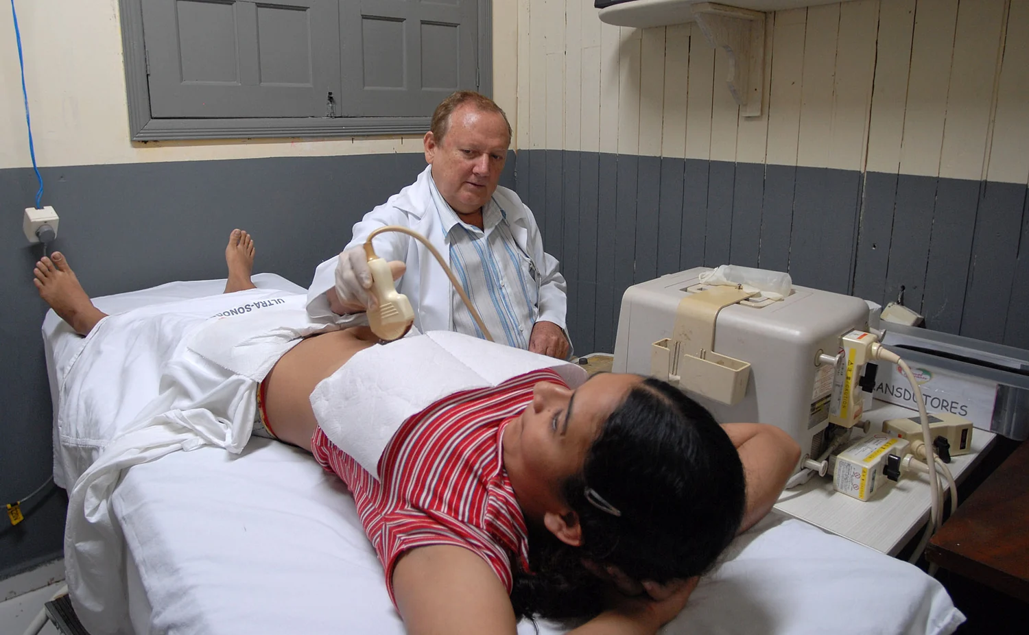
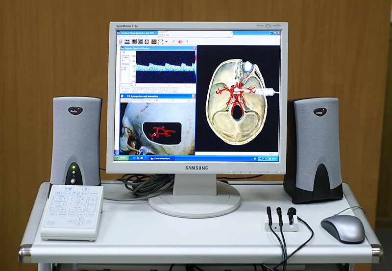
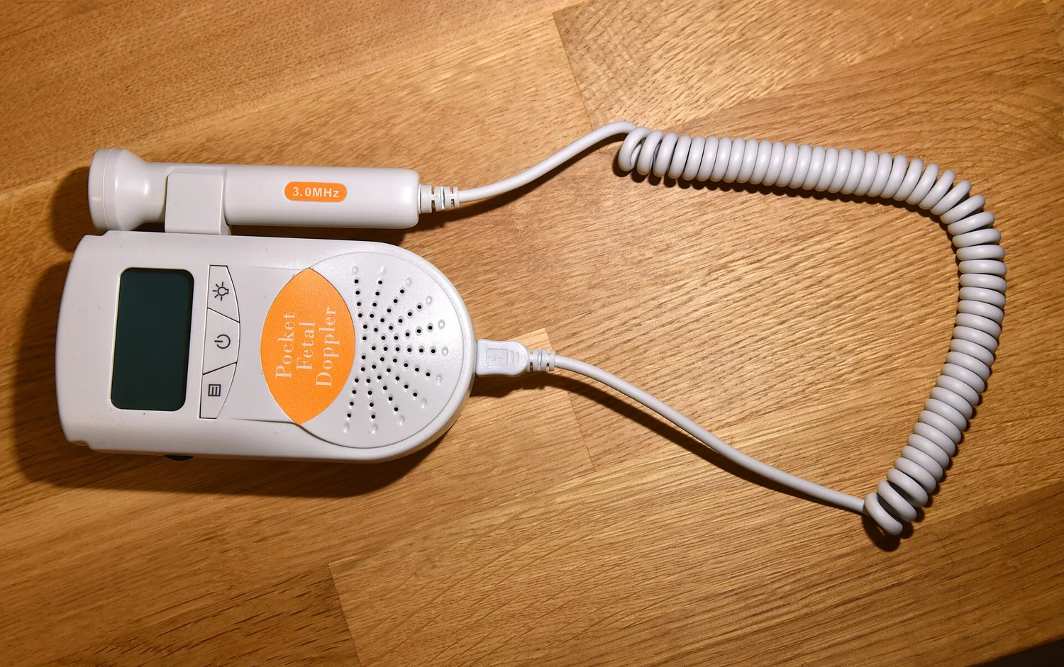
Ultrasound uses sound at frequencies above the threshold of human hearing, and diagnostic probes commonly operate in the megahertz range. A transducer sends sound into the body; boundaries between tissues reflect some of it back. The scanner times the returning echoes and uses the speed of sound to estimate the distance to each boundary [1]. This is a way to turn wave travel into an image.
Frequency matters because the wavelength of sound in tissue affects the scale of structures that can be distinguished. In general, shorter wavelengths can help resolve smaller details, while higher-frequency sound tends to be absorbed more as it travels, limiting how deeply it can reach. The practical choice is therefore a trade-off between detail and depth, not a contest to use the highest frequency available.
A medical signal can be sound or electromagnetic radiation
Ultrasound is a mechanical sound wave: it travels by vibrations passing through a material. Radio waves, visible light, and x-rays are electromagnetic radiation, consisting of changing electric and magnetic fields. They share the frequency–wavelength relationship, but their speeds and interactions with tissue differ. Ultrasound’s echoes arise at tissue boundaries; electromagnetic waves interact with matter according to their frequency and other properties.
The electromagnetic spectrum extends from long-wavelength, low-frequency fields to short-wavelength, high-frequency radiation. The National Cancer Institute distinguishes non-ionizing radiation, including radio waves, from ionizing radiation such as x-rays and gamma rays, which have enough energy to damage cells or DNA directly [2]. Frequency is one way to locate a signal on this spectrum, but it should not be mistaken for a complete description of its biological effect.
Frequency alone does not tell the whole story
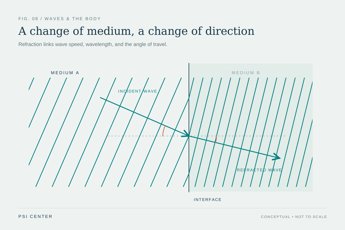
Two waves with different frequencies may travel at different speeds because they are different kinds of waves or move through different materials. Even for the same wave type, the surrounding medium can change speed and wavelength. In ultrasound, for example, the scanner uses an assumed speed of sound in tissue to interpret echo timing; boundaries and tissue properties influence what signal returns [1].
So when comparing medical signals, ask three questions: what is oscillating, where is the wave traveling, and how does the detector use the returned or received signal? Frequency tells how quickly the pattern repeats, wavelength tells its spatial spacing, and speed links the two. Those definitions offer a reliable starting point for understanding why an ultrasound probe, a radio transmitter, and an x-ray machine produce fundamentally different signals.
Explore the physics: an explanatory diagram
Sources & further reading
- Ultrasound NIBIB
- Electromagnetic Fields and Cancer National Cancer Institute
Prepared for the independent PSI Center reading library. About edition dates and source checking.
This explainer describes scientific principles. It does not assess individual treatment choices.