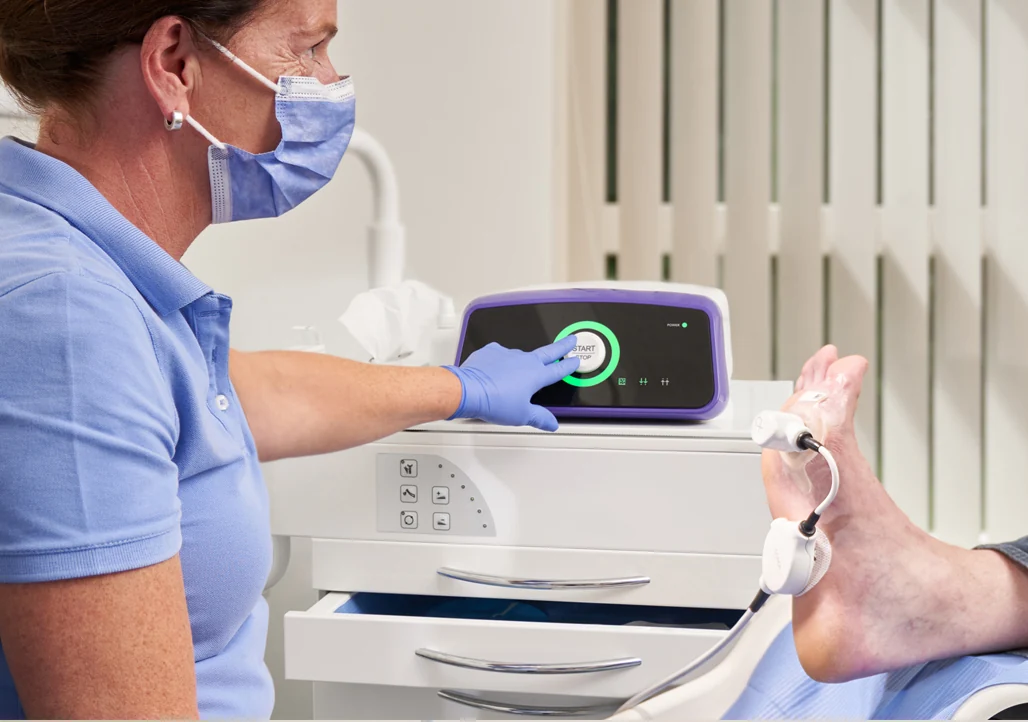
A small plasma jet may glow like a flame, but its medical interest comes from charged particles and reactive chemistry. Understanding the device also means distinguishing laboratory promise from established wound-care evidence.
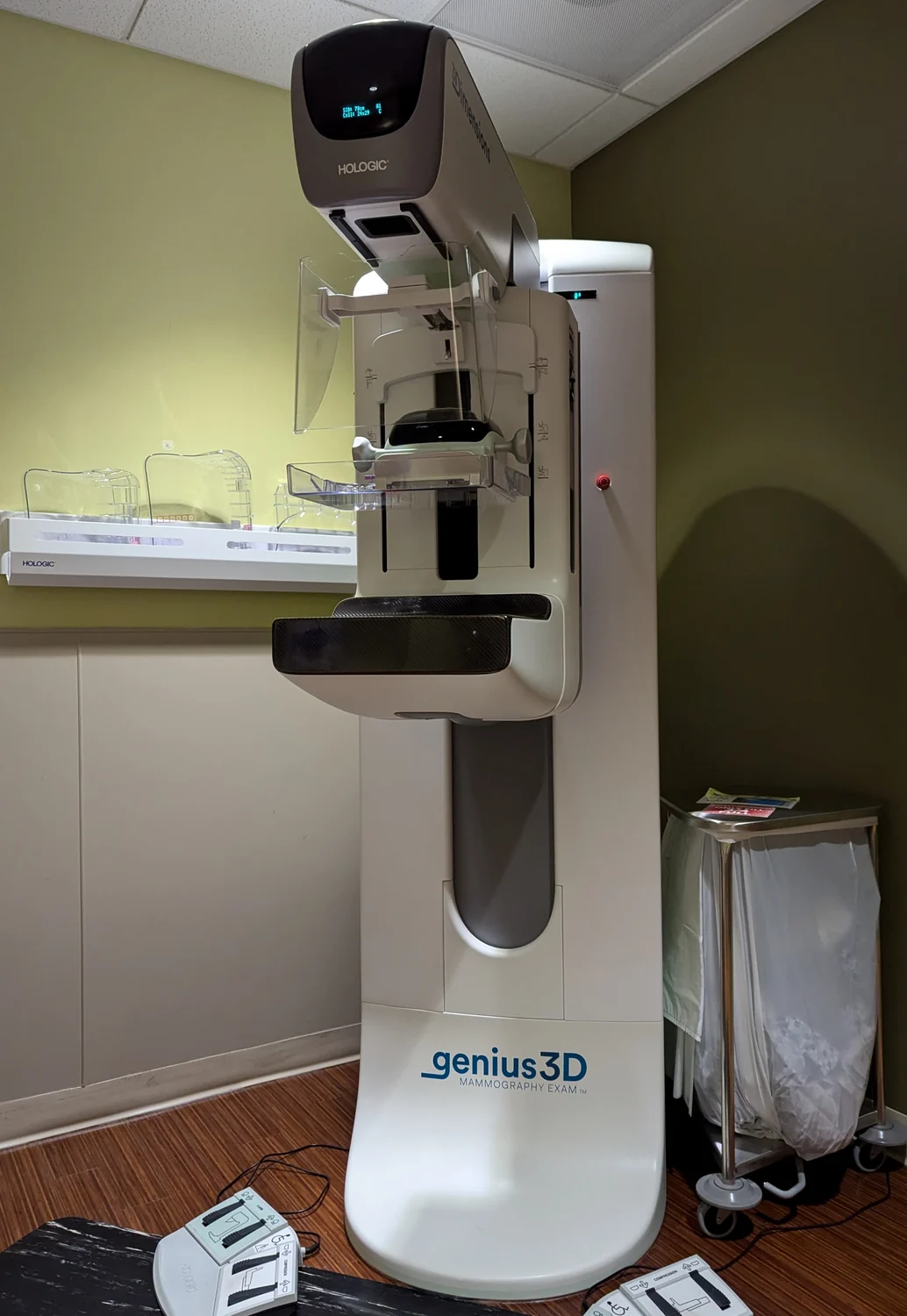
Mammography is radiography tailored to breast structures, with dedicated image receptors and quality requirements. Its aim and image geometry distinguish it from CT and live fluoroscopy, though all use ionizing X-rays.
A microscope turns light from a tissue sample into a magnified image that pathologists can inspect. The diagnostic value comes from how light interacts with structures and how image evidence is interpreted in clinical context.
Muscles produce electrical activity as nerves activate them. Surface sensors and needle electrodes sample that activity from different positions, so they answer related but nonidentical questions about nerve and muscle function.
A CT image is reconstructed from many x-ray measurements taken around the body. Computation turns those projections into slices, and a stack of slices can reveal how anatomy is arranged in three dimensions.
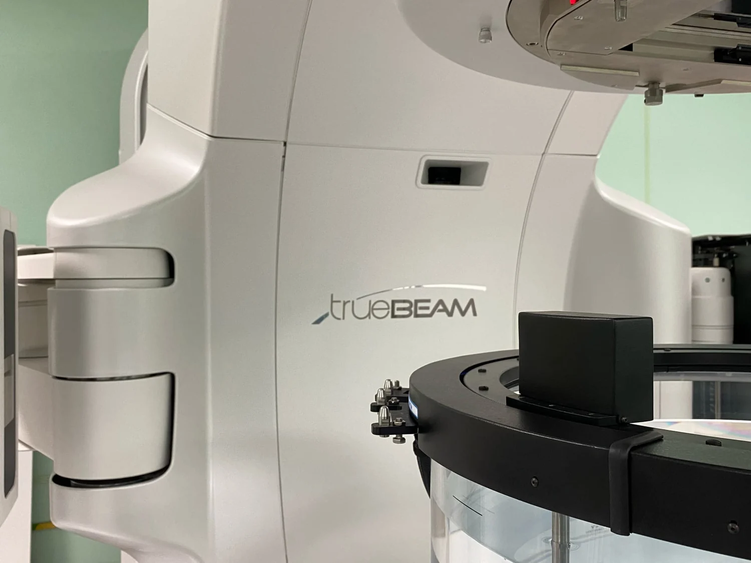
Radiation planning turns scans, beam properties, and a prescribed target into a calculated treatment plan. Modeling helps estimate where energy will go while accounting for the surrounding anatomy.
A dialysis membrane separates blood from dialysis solution while allowing selected substances and water to move across. Its usefulness comes from controlling exchange, not from acting as a simple sieve with identical pores.
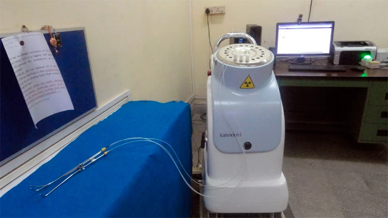
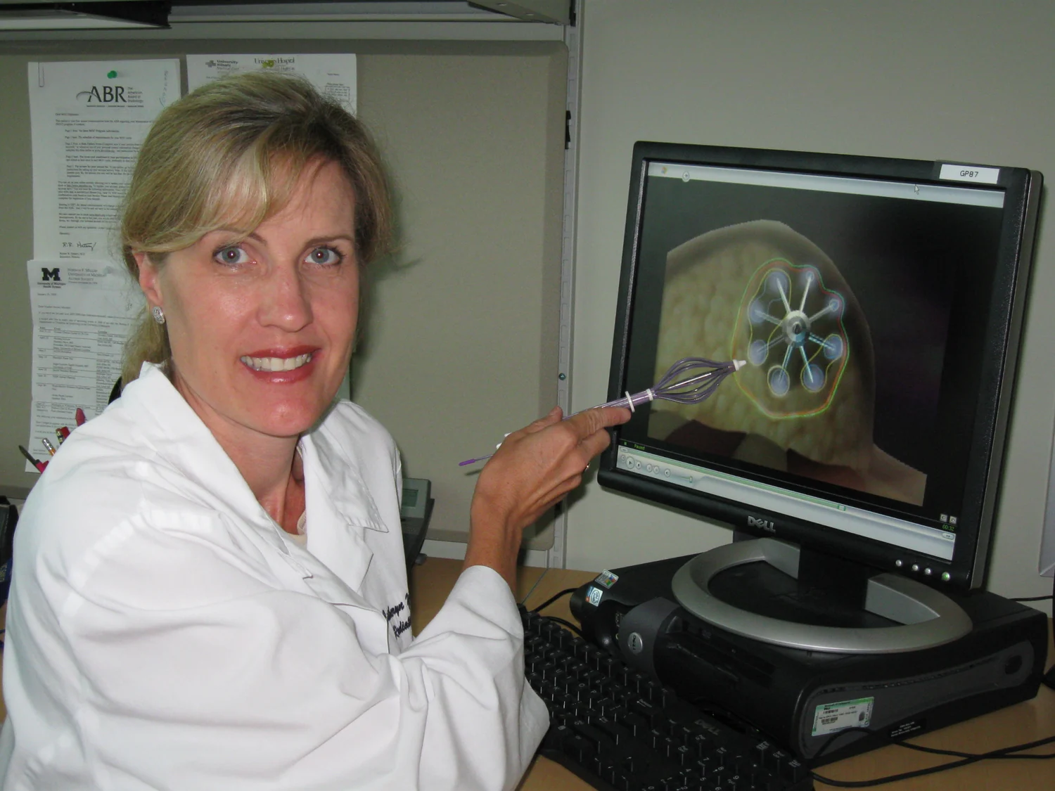
Brachytherapy places a radiation source in or near a tumor. Because radiation intensity and tissue exposure vary with geometry, source placement is central to delivering a local treatment while limiting exposure beyond the target.
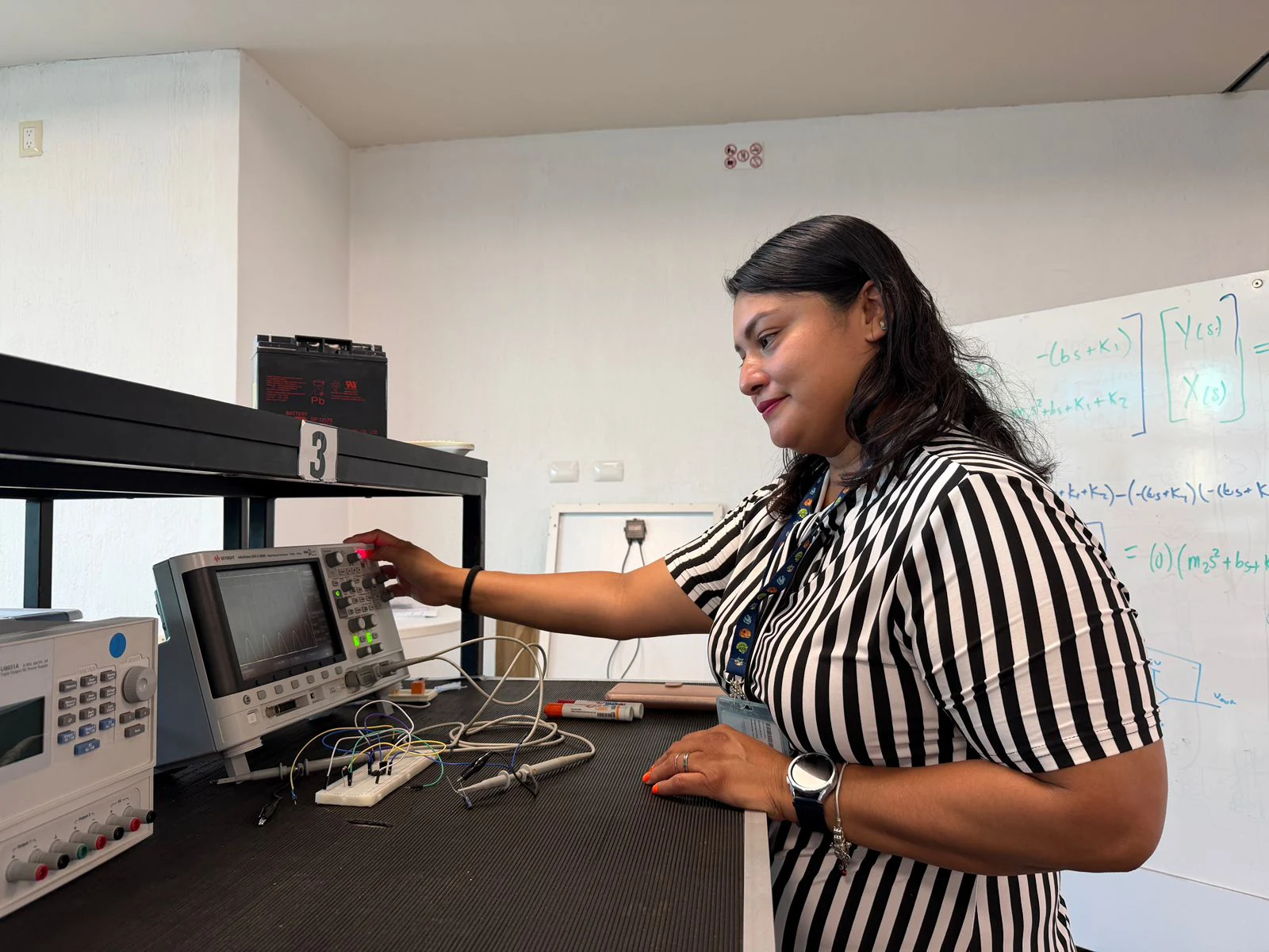
Electrodes record electrical differences at the skin, so contact quality affects how faithfully a small biological signal reaches the instrument. ECG and needle EMG illustrate two distinct ways of sensing activity.
A treatment can produce no obvious harm in a small study without proving it helps patients. Separating safety from effectiveness clarifies what early research can establish and what later trials must test.
A drug carrier can change where a medicine travels, how long it survives, and when it is released. The material’s chemistry and structure shape those effects, while biological complexity limits how precisely delivery can be controlled.
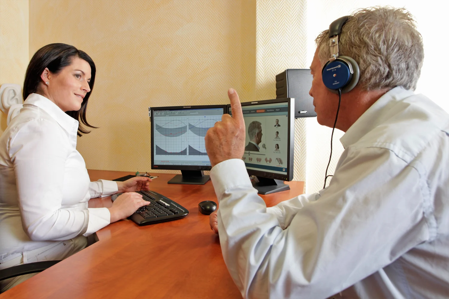
Decibels compress a vast range of sound pressures into a manageable scale. Understanding the logarithm behind the number helps explain why a small numerical increase can mean a large physical change—and why exposure time matters to hearing.
Proton therapy differs from photon radiation in how energy is deposited along a beam’s path. That physical distinction can help shape treatment, while clinical benefit depends on tumor geometry, nearby organs, and evidence for each cancer.
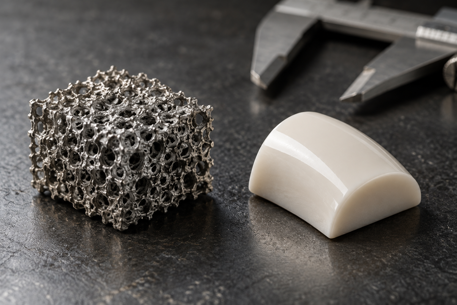
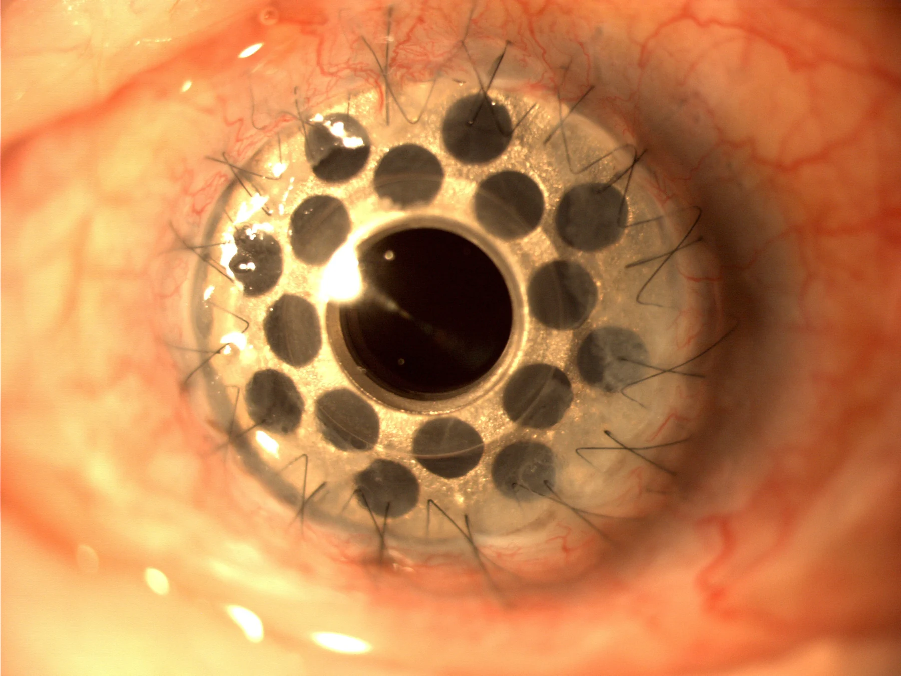
A coating can change the first material a cell encounters without changing the implant’s basic shape. Its chemistry and texture influence the interface, but a promising surface feature does not guarantee a clinical outcome.
Cold atmospheric plasma is an emerging wound-care approach, but device names alone reveal little about treatment delivery or evidence. Understanding trial design and outcome measures helps keep promising findings in proportion.
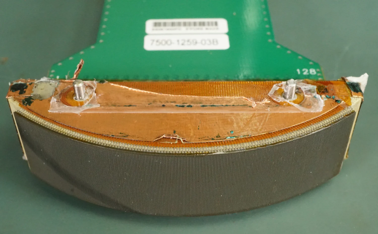
Diagnostic ultrasound sends sound pulses and listens for echoes to build images. Shock wave lithotripsy uses focused pressure waves to fragment kidney stones, turning acoustic energy toward a therapeutic task.
Models turn selected physical and biological details into testable predictions. Their value depends on clear assumptions, useful measurements, and knowing which questions the simplified system can—and cannot—answer.
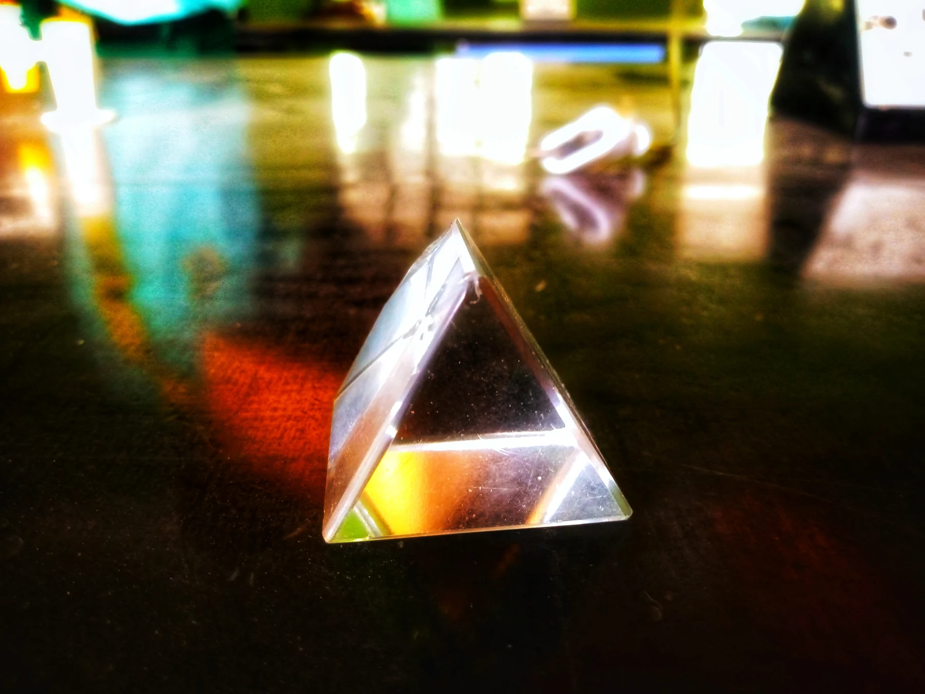
A photon’s energy depends on its wavelength because light comes in quantized packets whose energy tracks frequency. This link helps explain why ultraviolet light differs physically from radio waves and visible light.