What can an electrical signal recorded from a moving arm tell us about the nerves and muscles beneath the skin? Electromyography, or EMG, measures electrical activity associated with muscle action. The location of the electrode changes what the instrument can detect: a sensor on the skin samples a broad, mixed signal, while a needle electrode sits within a selected muscle. That difference shapes what each measurement can reveal.
Muscle activity has an electrical footprint
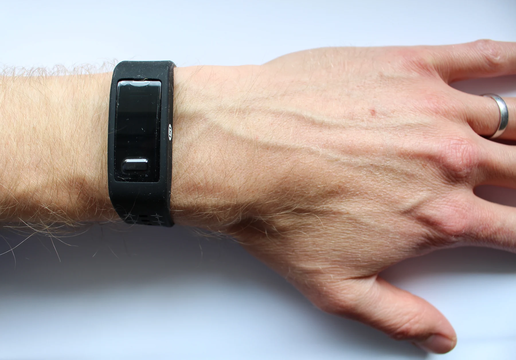
A muscle contracts when nerve signals activate muscle fibers. The resulting electrical changes spread through tissue and can be measured at the body surface or closer to their source. A sensor is a device that detects a physical or biological input and turns it into a reportable signal; in biomedical settings, sensors may report electrical, chemical, mechanical, or optical changes [2].
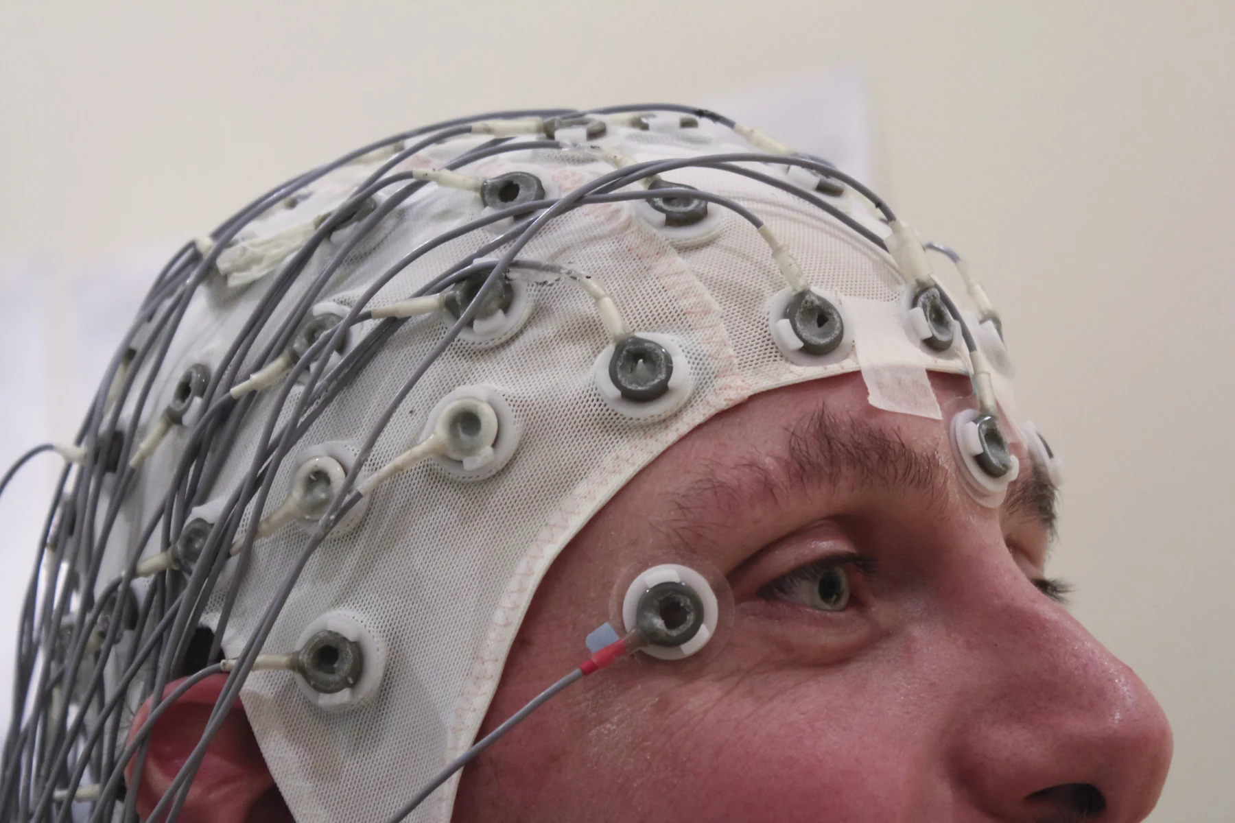
Surface electrodes placed on the skin can record activity without entering the muscle. Their signal combines contributions from many fibers and is affected by distance, skin contact, movement, and electrical activity in neighboring muscles. This makes surface recording useful for observing patterns across time or during movement, but it is not a direct view of each individual muscle fiber. The sensor reports what reaches it through the intervening tissues.
Why place a needle electrode?
A clinical needle EMG uses a very thin electrode inserted through the skin into a muscle. Because it sits closer to active fibers, it can pick up electrical activity from a more localized area. The examiner may record the muscle at rest and ask the person to contract it, then observe how the signal changes. Normally there is little activity in a relaxed muscle, while contraction produces increasing activity and a pattern that can be assessed [1].
The needle approach is invasive in a limited, specific sense: it crosses the skin to sample inside a muscle. It may cause brief discomfort, and the area can feel tender or bruised afterward. Reported risks include minimal bleeding and rare infection at electrode sites [1]. Those practical tradeoffs differ from a skin electrode’s, but the purpose is not simply to collect a “better” signal. It is to sample from a more specific location when that information is useful.
Two measurements, two views
Consider a person asked to bend an arm. A surface electrode may register a changing electrical pattern from muscles under and near the sensor. A needle electrode placed in one muscle records local activity there, including whether activity appears at rest or changes during contraction. The contrast resembles listening to a room with a microphone versus placing a microphone near one instrument: both record real sound, but they mix different sources.
EMG is often performed alongside a nerve conduction velocity test. That test measures how fast and how strongly electrical signals travel along a nerve; it complements muscle recording by assessing the nerve pathway itself [1]. Together, these measurements can help distinguish weakness associated with nerve injury from weakness related to muscle or nervous-system disease. They are not interchangeable: a muscle signal and a nerve-conduction measurement describe different links in the system.
Reading a pattern, not a label
An EMG result is interpreted as a pattern in context. Electrical activity may be unusual at rest or during contraction, and abnormal patterns can occur across many conditions, including nerve disorders and muscle diseases. The test can help investigate weakness, pain, or altered sensation, but a signal alone does not identify every cause. The clinician considers which muscles were sampled, the examination, and the accompanying nerve-conduction findings [1].
Sensors are designed around the quantity they detect, and not every sensor is a diagnostic instrument. Wearable technologies can track measures such as heart rate or sleep, while some advanced sensors remain research tools rather than routine-care devices [2]. The same principle applies to EMG: surface and needle electrodes are not competing versions of one universal test. Each samples a different part of the electrical picture. Knowing where the electrode sits helps explain both the detail it can provide and the limits of what its trace means.
Sources & further reading
- Electromyography MedlinePlus
- Sensors NIBIB
Prepared for the independent PSI Center reading library. About edition dates and source checking.
This explainer describes scientific principles. It does not assess individual treatment choices.