How can metal disks on the scalp record activity from cells inside the brain? They do not listen to individual neurons. An electroencephalogram, or EEG, measures small voltage differences that reach the scalp from many brain cells acting together. The resulting wavy traces change with sleep and alertness and can help clinicians evaluate neurological conditions, but they are signals to interpret—not a direct picture of thoughts or a complete record of every event.
From brain impulses to scalp voltage
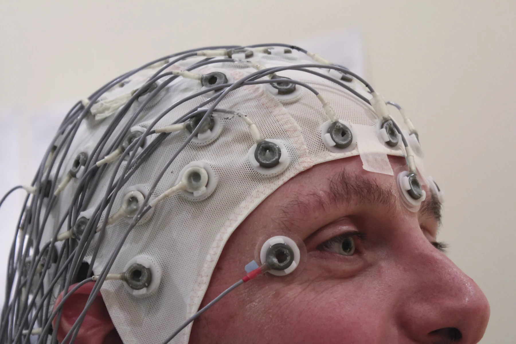
Brain cells communicate through tiny electrical signals, or impulses. When many cells’ activity is coordinated, some of the resulting electrical fields can be detected at the scalp. EEG electrodes are flat metal disks held in place with paste; wires connect them to a recording device that turns changing signals into patterns on a screen or paper [1].
The scalp is not a transparent window. Signals from deeper sources spread through brain tissue, fluid, skull, and skin before reaching an electrode, so the measurement is spatially blurred. A useful analogy is hearing a crowd from outside a hall: the sound level reveals changing group activity, but not the words of each person. EEG records collective electrical patterns, not a neuron-by-neuron map.
Why the trace changes
EEG waves vary with brain state. For example, waves are faster during wakefulness and slower during some stages of sleep. A recording session may include quiet rest, sleep, or tasks such as deep breathing or looking at flashing light, because changing the state or stimulus can reveal patterns relevant to the clinical question [1].
Movement can alter the trace, which is why people are generally asked to lie still during a routine test. Blinks, muscle activity, and other electrical sources can also contribute signals at the scalp. This is one reason EEG interpretation depends on the pattern, timing, and recording conditions rather than treating every wiggle as a brain event.
A recording matched to a question
EEG can be used to diagnose or monitor seizures and epilepsy, sleep disorders, certain brain diseases, coma, unexplained fainting or memory loss, and other conditions. It can also monitor brain activity during surgery. A normal result does not prove that a seizure did not occur: the test samples activity during a particular recording window, and not every event appears in that window [1].
If longer monitoring is needed, ambulatory EEG can record activity while a person follows a normal routine, or monitoring may occur in a hospital unit. Video may be recorded alongside electrical signals so that clinicians can compare what the person was doing with the EEG pattern at that moment [1].
A sensor is not a mind reader
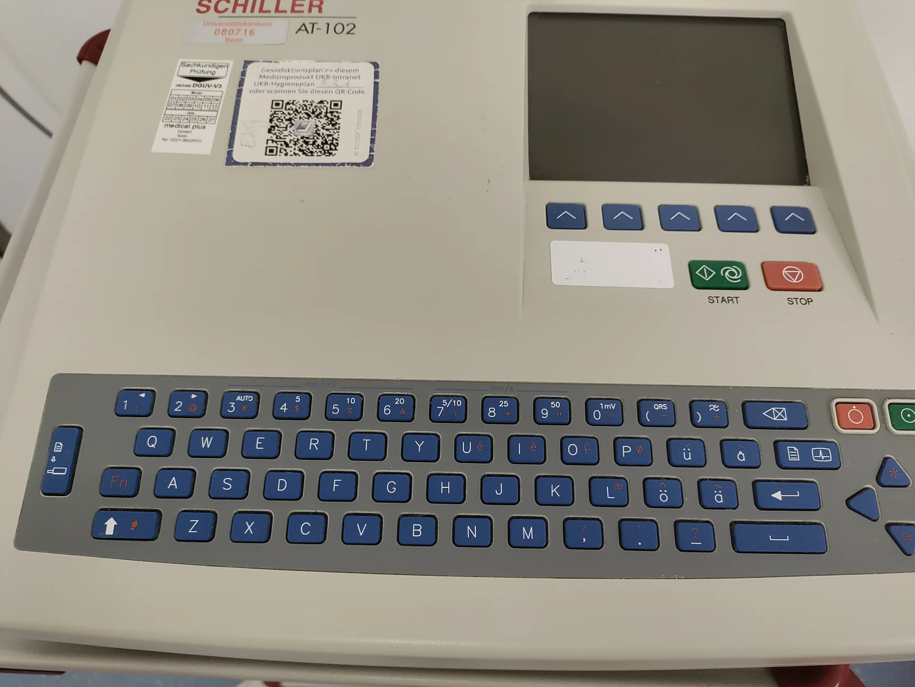
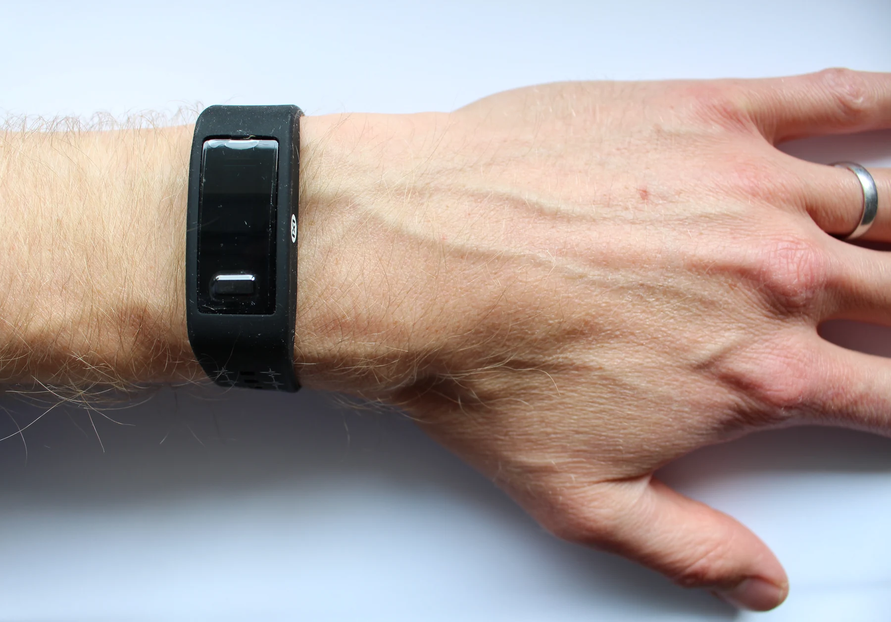
A sensor detects and responds to an input from its environment. Biomedical sensors may detect electrical, optical, chemical, pressure, or temperature signals, then report data to a user or health professional [2]. EEG is one sensor system: its electrodes detect scalp voltage changes, and its recording equipment displays them in a useful form. The displayed waves are measurements, not a direct reading of a person’s experience.
EEG cannot be used to measure intelligence [1]. Nor does every unusual-looking result point to one cause: abnormal patterns may be associated with multiple conditions or factors, including medicines, injury, infection, or seizure disorders. The test is most informative when its limits are kept in view: it records electrical activity over time, and clinicians interpret that evidence alongside the reason for testing and other information.
Sources & further reading
- Electroencephalogram MedlinePlus
- Sensors NIBIB
Prepared for the independent PSI Center reading library. About edition dates and source checking.
This explainer describes scientific principles. It does not assess individual treatment choices.