Why can a sensor on the skin record a heartbeat, yet a loose or poorly placed electrode make the trace harder to read? The electrode does not reach into the heart; it senses tiny voltage differences conducted through body tissues. The quality of the electrical contact helps determine how much of that signal reaches the recording system and how much unwanted variation joins it. Comparing an ECG sticker with a needle electrode shows how sensor placement shapes what can be measured.
The body produces signals; electrodes detect them
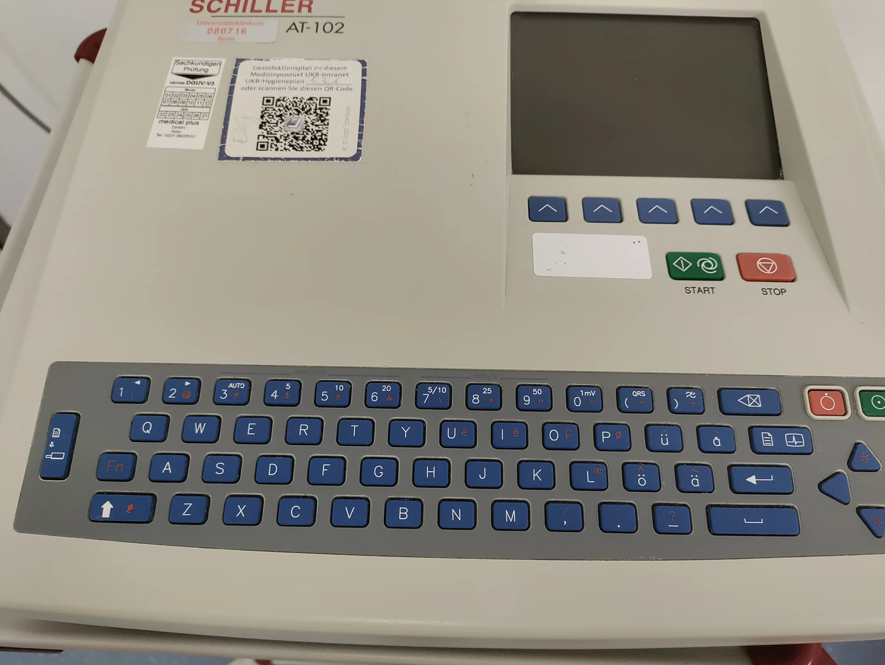
An electrocardiogram, or ECG, records electrical signals associated with the heart’s beating. Those signals coordinate chamber contractions, and the recorded pattern appears as waves with recognizable parts such as the P wave, QRS complex, and T wave [1]. The ECG machine does not send electricity into the body; it records signals through electrodes placed on the skin [1].
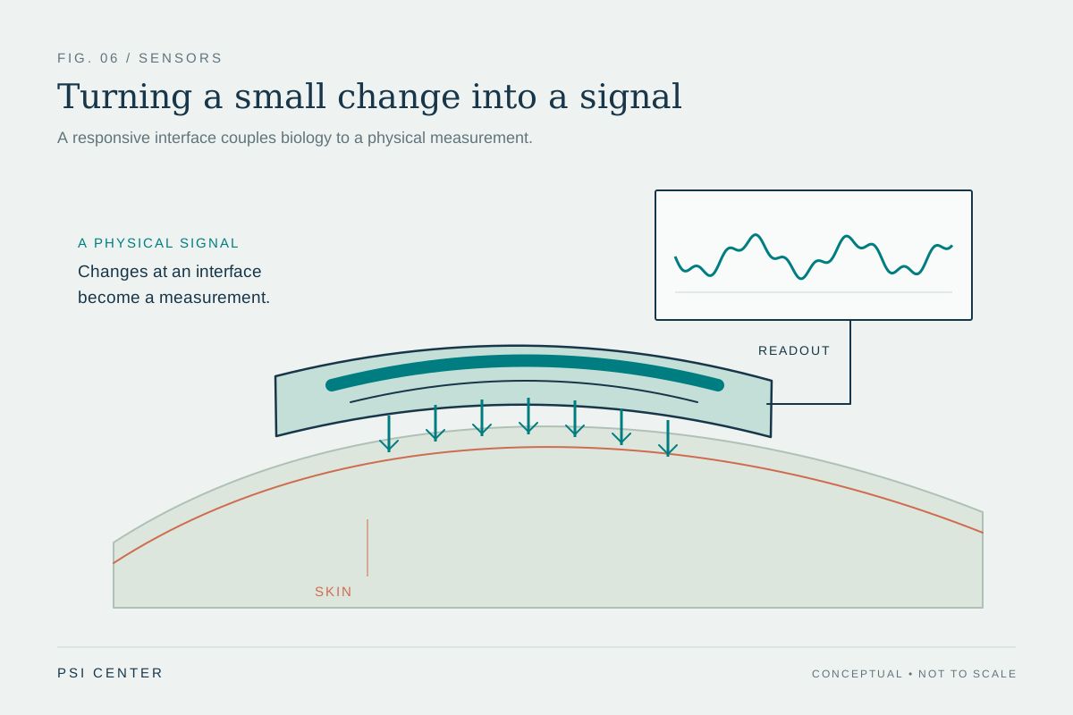
The heart’s electrical activity spreads through conductive tissues before reaching the skin. A recording system compares voltage at different electrode locations, so the measured trace depends on both the source and the path from body to sensor. Contact is the interface in that path. If it is inconsistent, the signal may be less stable or more vulnerable to movement and electrical interference.
Contact is an electrical boundary
A sensor touching skin meets a surface that is not a perfect, uniform conductor. Hair, dry skin, movement, and gaps can change the electrical connection. Better, steadier contact makes it easier for the instrument to measure the voltage differences it is designed to record. This is why ECG electrodes are made to stick to the skin and may require preparation to stay in place [1].
Think of a microphone cable with a loose plug: the sound source has not changed, but the connection can add crackle or drop parts of the signal. A biological electrode is not a microphone, and its signal is electrical rather than acoustic, but the analogy captures the role of the interface. Poor contact can distort the measurement without implying that the heart itself has changed.
Different electrodes answer different questions
A surface ECG samples electrical activity from the heart as it reaches the skin. It can show rate, rhythm, and the timing and strength of signals across parts of the heart, but the recording is a snapshot and cannot diagnose a condition by itself [1]. If symptoms come and go, longer monitoring may capture activity missed during a brief test [1].
Electromyography, or EMG, uses a different sensor and asks a different question. A very thin needle electrode is inserted into a muscle to pick up its electrical activity at rest and during contraction; the pattern helps assess how muscle responds and can help distinguish some nerve-related weakness from muscle or nervous-system disease [2]. The needle’s location samples activity inside a particular muscle, unlike skin electrodes that record signals across the body surface.
Signal quality is not the same as diagnosis
In an EMG, activity typically rises as a person contracts the sampled muscle, and the observed pattern is interpreted in the context of the test [2]. The instrument turns electrical changes into a display or sound, but the display is evidence to be interpreted, not a diagnosis by itself. The same principle applies to ECG: an unusual trace can be a clue, while clinical meaning depends on other information and evaluation [1].
Electrode contact matters because measurement starts at an interface. Stable contact helps preserve the signal; placement determines which parts of the body contribute; and the sensor type determines what scale of activity is sampled. Surface ECG and needle EMG therefore cannot be treated as interchangeable readings. Each is designed to capture a different electrical view of the body, with its own limits and interpretive context.
Explore the physics: an explanatory diagram
Sources & further reading
- Electrocardiogram MedlinePlus
- Electromyography MedlinePlus
Prepared for the independent PSI Center reading library. About edition dates and source checking.
This explainer describes scientific principles. It does not assess individual treatment choices.