What happens to a radiation beam after it reaches a tumor? A photon beam continues onward, depositing some energy in tissues along its route and beyond its target. A proton beam can instead be arranged to release much of its energy near a selected depth and then stop. That difference makes proton therapy physically distinctive, but it does not automatically make it the better treatment for every tumor.
Two kinds of beam, two paths through tissue
Radiation therapy uses high doses of radiation to damage the DNA of cancer cells, slowing their growth or killing them. The injured cells do not disappear instantly: their ability to divide fails, and cell death and removal can unfold over days, weeks, or longer after treatment [2].
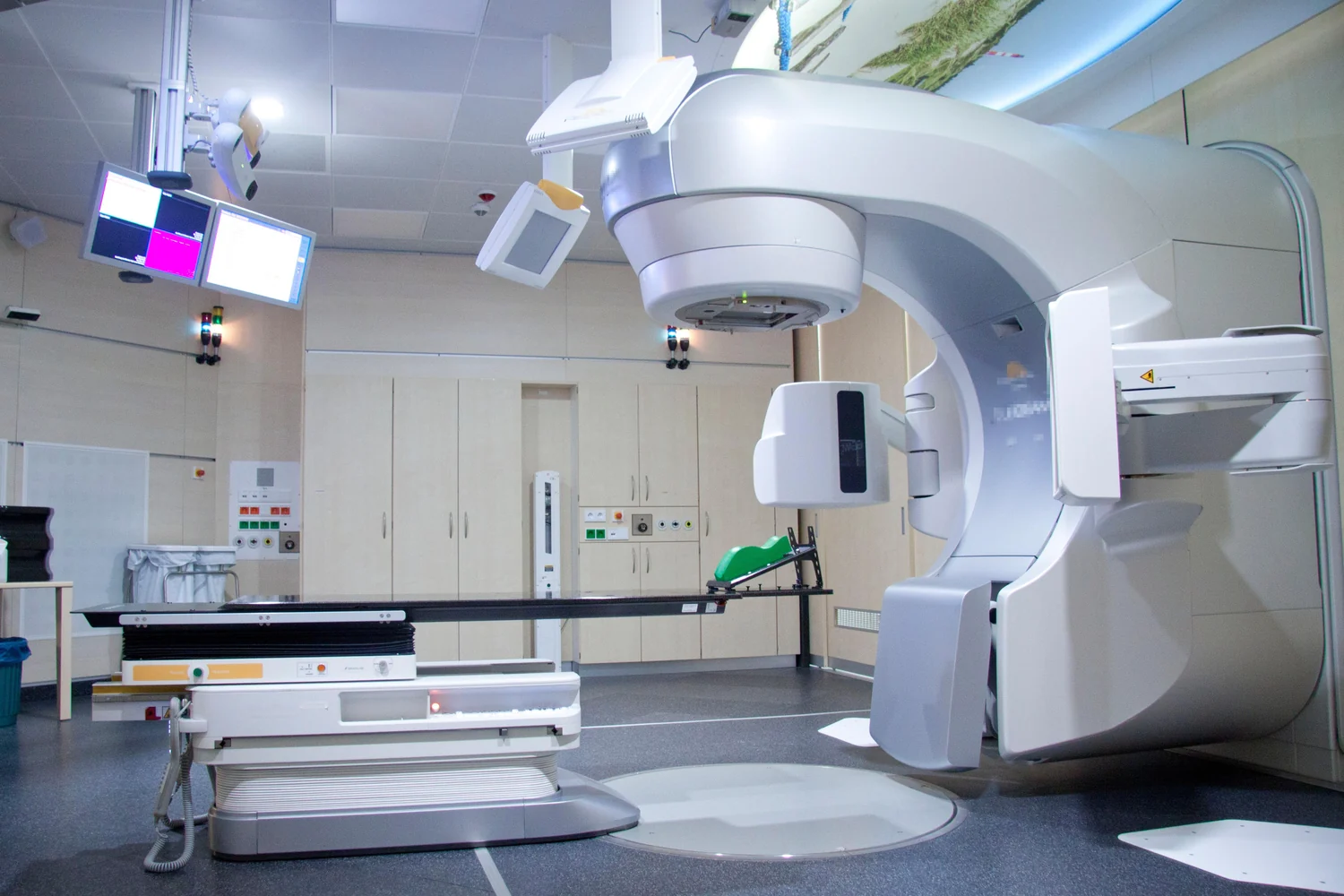
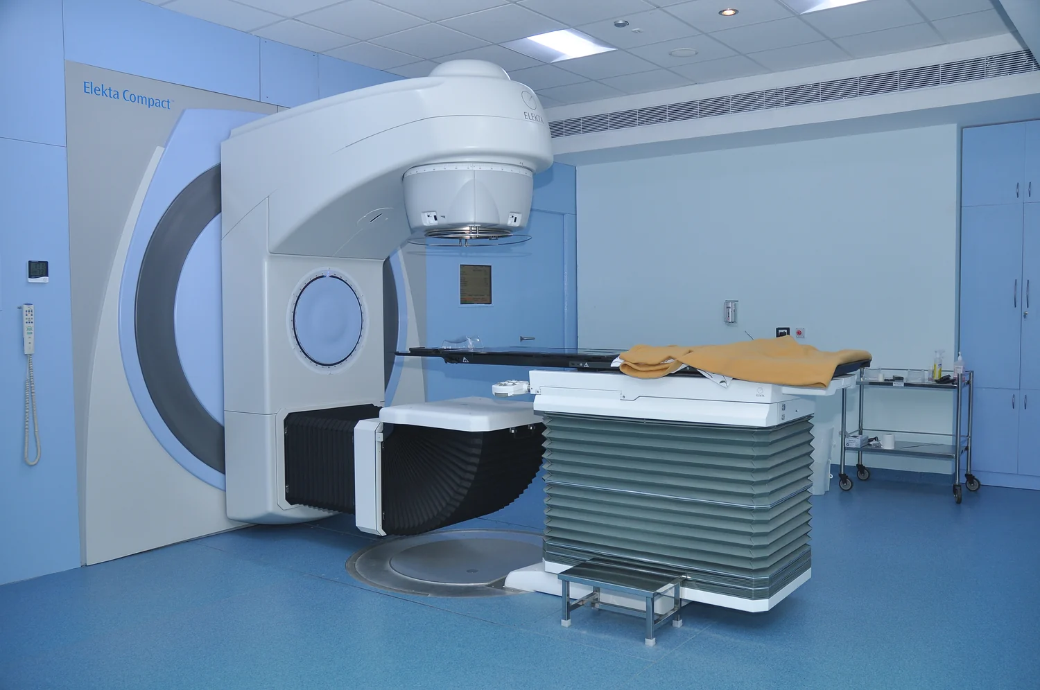
Most external radiation machines use photons, the same broad family of electromagnetic radiation that includes x-rays. A photon beam can reach deep structures, but it deposits energy as it travels and does not stop at the tumor. Some dose therefore reaches normal tissue both before and after the target. Protons are positively charged particles. Their path through matter is different: they can travel to depth and deposit much of their dose near the end of their range, with little dose beyond that point [1].
The depth effect: a useful mental picture
Imagine aiming two narrow streams toward a small target embedded in a block of tissue. One stream leaves a faint trace along the whole route and continues out the far side. The other leaves a trace on its approach, grows more intense near a chosen depth, and then largely ends. This is a simplified picture of photon and proton dose patterns; actual treatment planning uses detailed calculations, not a literal sharp boundary.
The proton’s concentrated end-of-range energy deposition is often called the Bragg peak. Adjusting particle energy changes how deep that peak falls. Combining energies and shaping beams can cover a tumor’s thickness. The practical opportunity is to reduce exposure to some healthy tissue beyond the tumor, or to sensitive tissue nearby, when geometry permits. It is not a promise that all normal tissue is spared: tissue before the target still lies in the beam’s path, and real beams have finite spread and range uncertainty.
Precision still depends on anatomy and motion
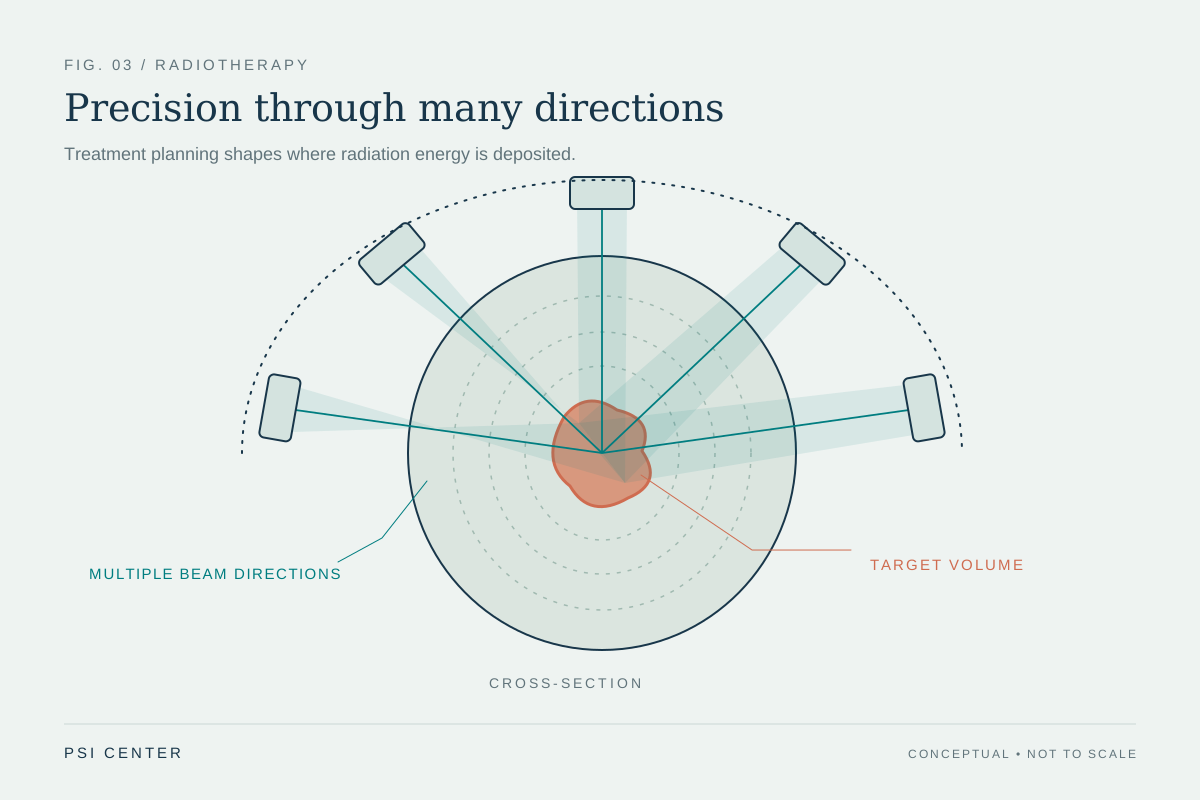
A tumor is not a stationary dot on a diagram. Organs shift with breathing, digestion, posture, and day-to-day changes. Planning therefore uses medical images and computer calculations to select beam paths and dose distributions. External beam treatments more generally aim to deliver the prescribed dose to the tumor while sparing surrounding tissue; image-guided methods can use scans during sessions to detect changes in position and adjust treatment [1].
The same anatomical details that make a proton plan attractive can also make it demanding. The particle must stop at the intended depth, so changes in tissue thickness or density along its route can alter where energy is deposited. A clinician’s planning question is not merely whether protons stop, but whether that stopping pattern can be predicted and maintained for the particular tumor and surrounding structures.
Physical advantage is not the same as proven outcome
A more favorable dose map is a physical result; a better clinical outcome is a separate question. Reducing dose to normal tissue could matter if it lowers side effects or permits a treatment plan that better serves a patient’s situation. But demonstrating such benefit requires clinical comparisons that measure outcomes, not just simulations or attractive images. The National Cancer Institute notes that clinicians think proton beams might reduce normal-tissue exposure and that clinical trials compare proton and photon approaches [1].
The choice of radiation approach depends on cancer type, tumor size and location, nearby radiation-sensitive tissues, health, and other treatments [2]. Proton facilities also require large, costly equipment, which limits availability [1]. Proton therapy is therefore best understood as a different tool for shaping where radiation goes. Its physical distinction is real; whether that distinction improves the balance of benefit and harm is a clinical question for each setting.
Explore the physics: an explanatory diagram
Sources & further reading
- External Beam Radiation Therapy National Cancer Institute
- Radiation Therapy to Treat Cancer National Cancer Institute
Prepared for the independent PSI Center reading library. About edition dates and source checking.
This explainer describes scientific principles. It does not assess individual treatment choices.