Interim findings from a randomized study of cold plasma and chronic lower-leg wounds offer a research signal, but their limits and a separate guideline recommendation shape what can be concluded for diabetic foot ulcers.
A 3D ultrasound display is assembled from sound echoes sampled across space. Transducer position, timing, and computer processing turn those measurements into a volume, whose appearance depends on the data collected.
Ultraviolet light is invisible, but its wavelength and energy determine how it interacts with skin. Sunscreen changes the radiation reaching tissue, while SPF and broad-spectrum labels describe different parts of protection.
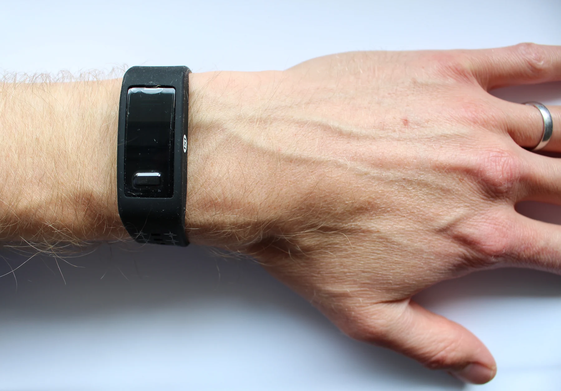
A pulse oximeter estimates oxygen saturation from changing light absorption in blood. Movement can scramble the small pulse-linked signal, while circulation, skin pigment, and sensor design also influence accuracy.
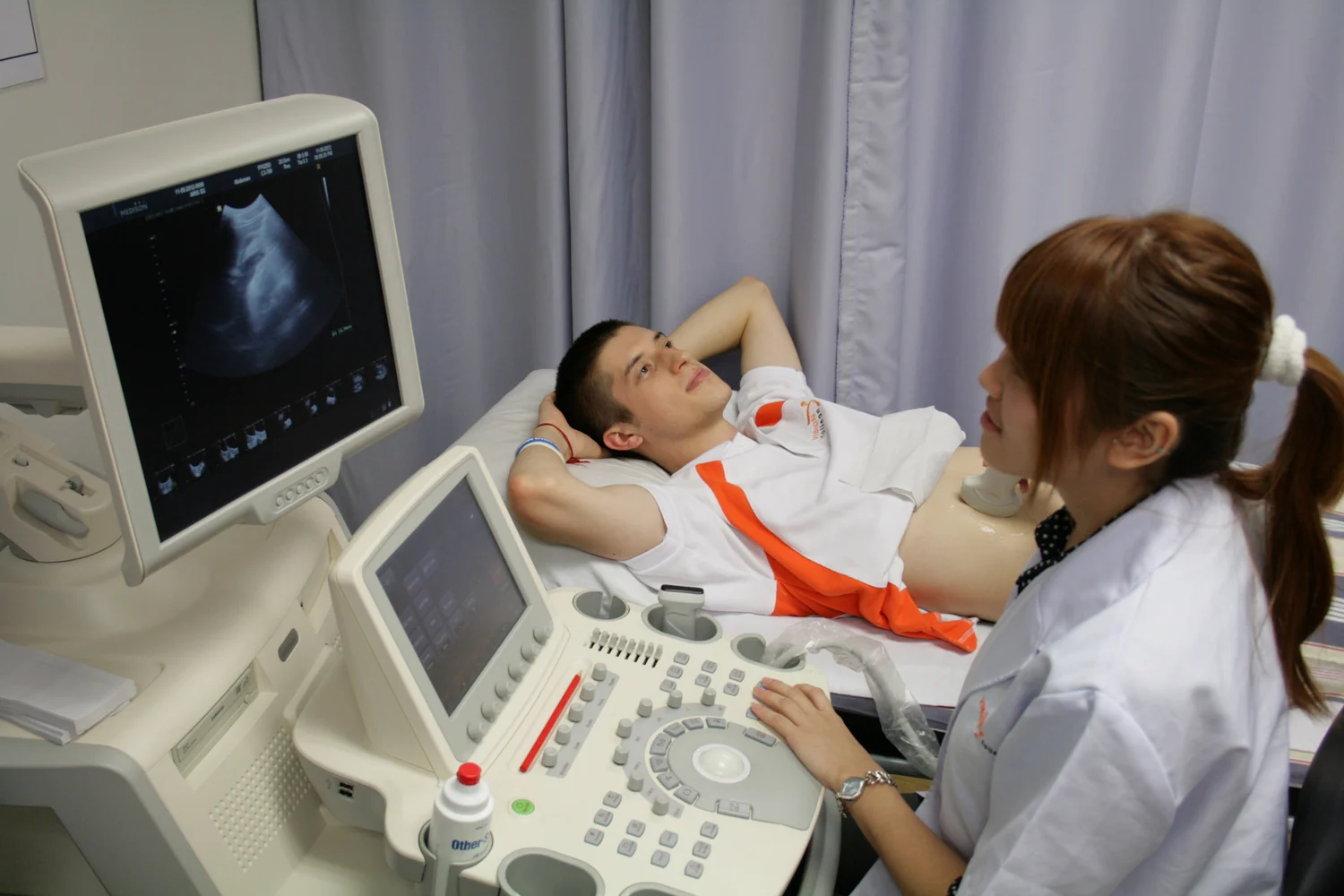
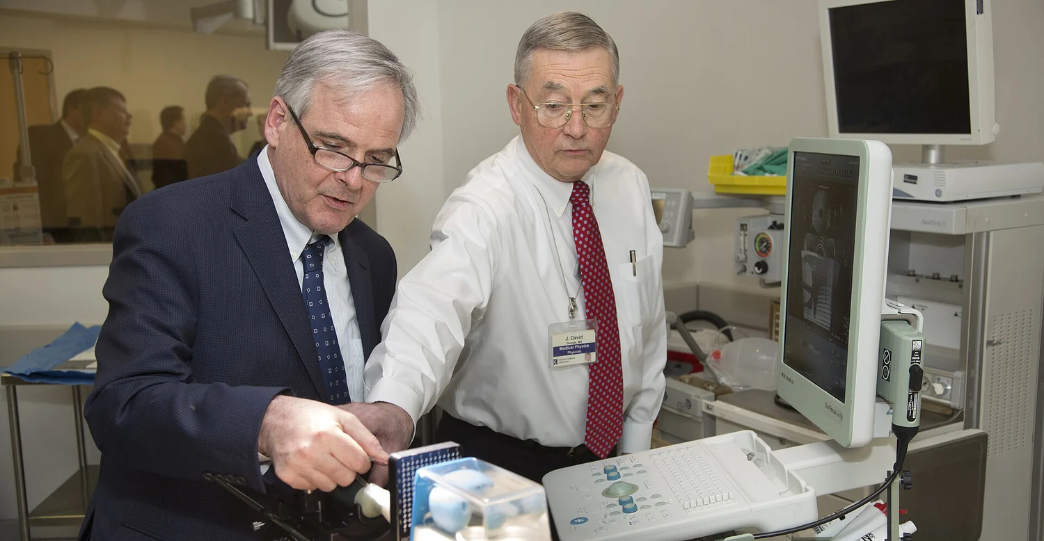
Ultrasound turns returning echoes into a live view of internal structures and motion. Its portability and real-time images can support assessment and guidance, while physics limits what the beam can see.
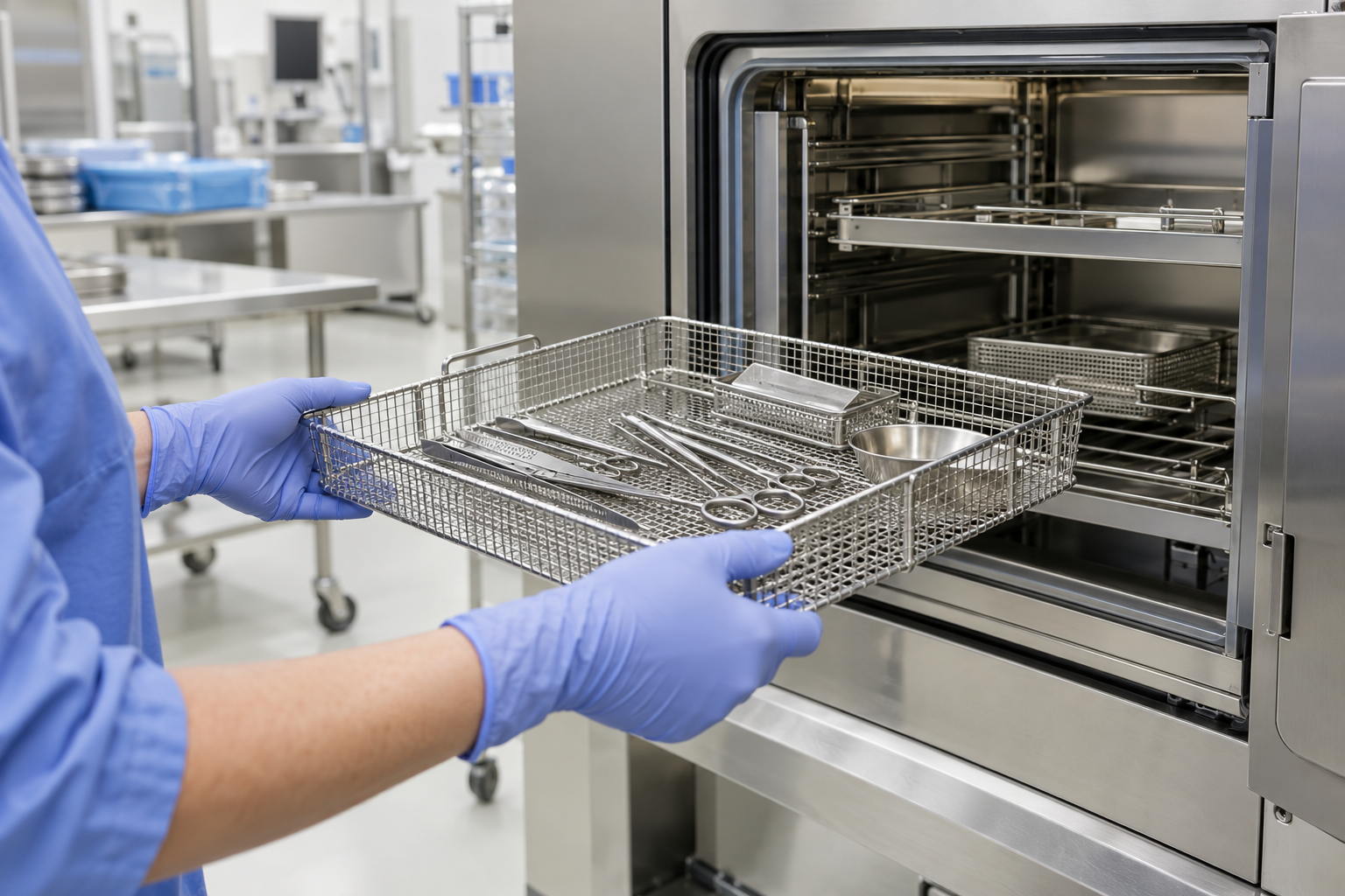
A sterile instrument is not certified by proving that every microorganism is absent. Sterility assurance estimates process reliability, which depends on cleaning, packaging, cycle selection, monitoring, and careful handling.
A quick test is the visible end of a longer chain: collecting a sample, converting biological signals into a result, and deciding what that result can support. Understanding each link clarifies both the value and limits of testing near the patient.
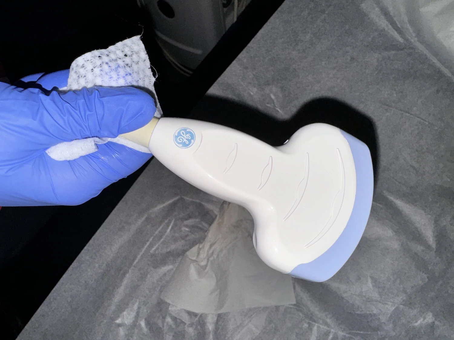
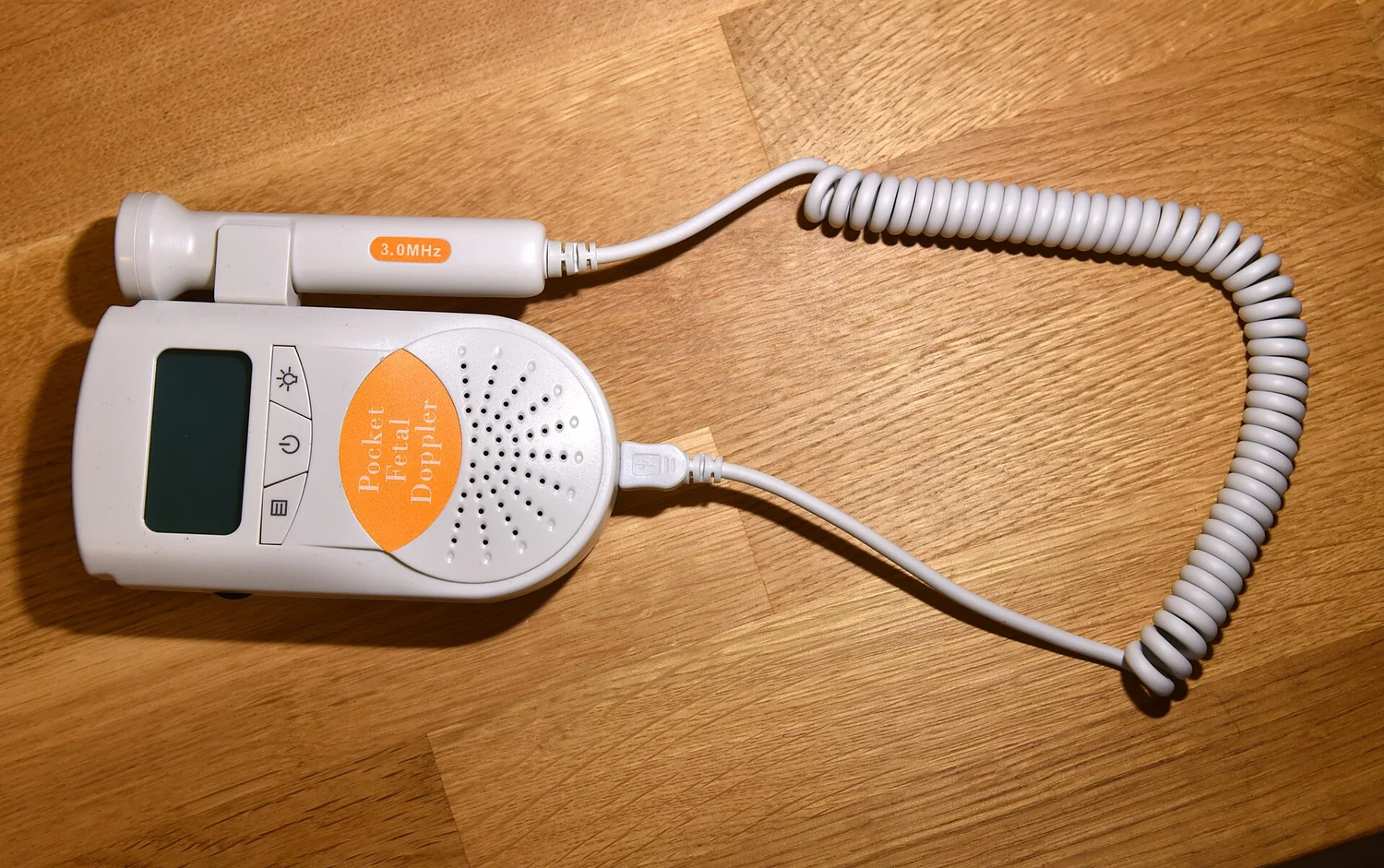
A probe turns electrical pulses into sound and returning echoes back into electrical signals. Their travel times and strengths help a scanner map tissue boundaries, while gel keeps air from blocking the path between skin and probe.
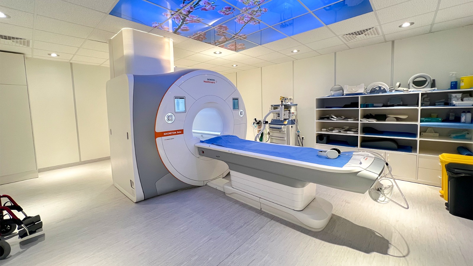
Electric and magnetic fields are related but arise in different ways and interact with matter differently. Their distinctions help explain medical technologies such as MRI and clarify why field exposure is not the same as ionizing radiation.
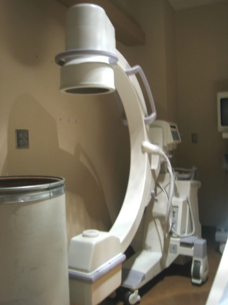
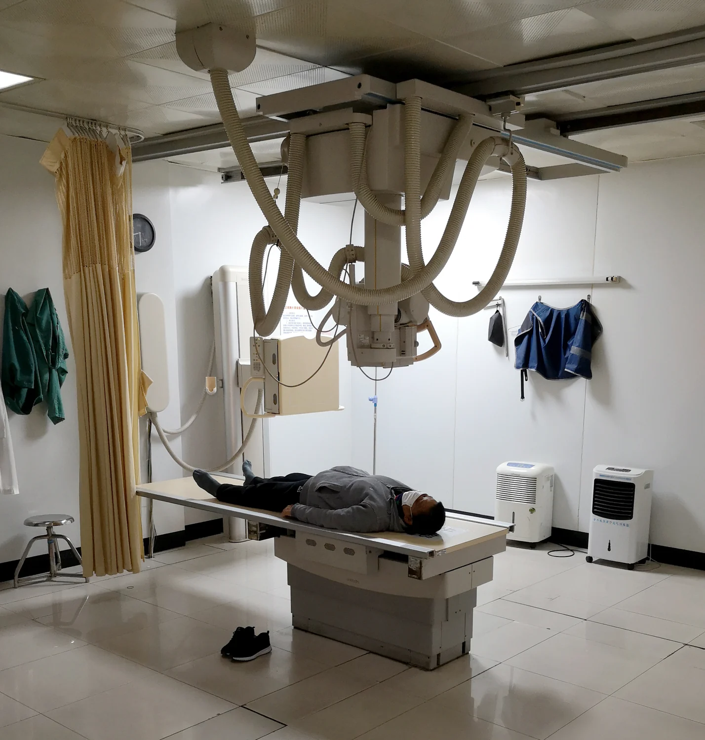
Fluoroscopy creates live images with X-rays, so exposure accumulates during a procedure. Time is one part of dose management, alongside clinical need, image quality, patient factors, and optimized technique.
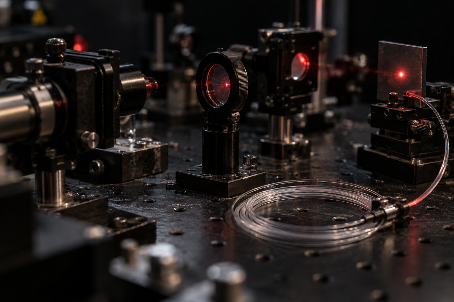
The eye’s focusing system can concentrate a narrow laser beam onto light-sensitive tissue. Understanding that optical path explains why exposure controls depend on the beam, its class, and how it is viewed.
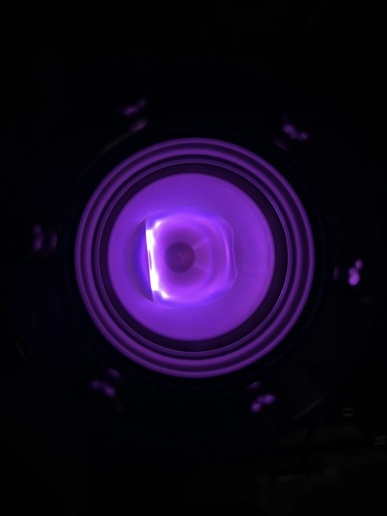
Free electrons help make a plasma electrically responsive and chemically reactive. Those features underpin distinct medical uses, from experimental interaction with living tissue to hydrogen peroxide plasma sterilization of equipment.
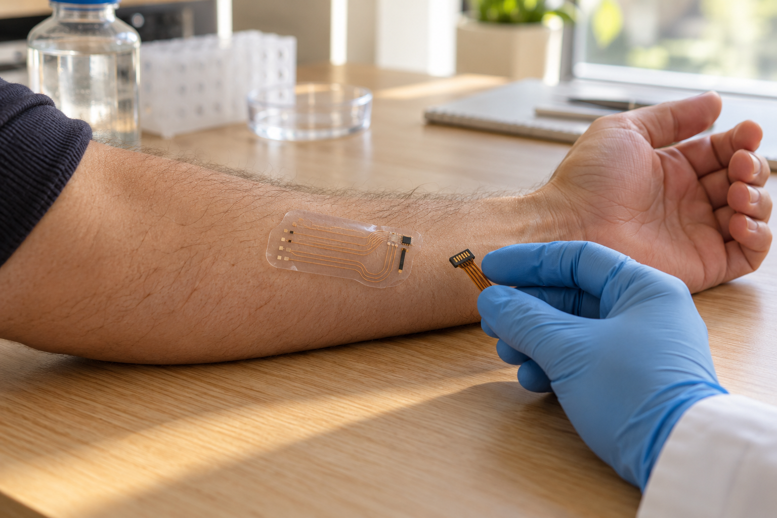
Repeated sensor readings can cluster tightly yet sit away from the true value. Separating precision from accuracy clarifies what a device reports and what its pattern of error means for interpretation.
Cancer research moves through questions of mechanism, safety, and clinical benefit. Keeping those stages distinct helps explain what a promising result can—and cannot—tell patients.
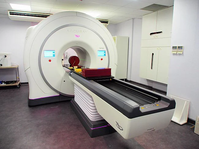
Radiation treatment depends on delivering a planned dose to a defined target while limiting exposure to nearby tissue. Measurement chains and repeated imaging help make those plans interpretable and checkable.
Sterilizing a medical device means controlling microbes without sacrificing the material’s function. Hydrogen peroxide gas plasma can suit some heat-sensitive polymers, but compatibility depends on device design and process conditions.
Electromyography records electrical activity in muscle, while nerve conduction testing measures how signals travel along nerves. Together, these measurements can help distinguish patterns of nerve-related and muscle-related weakness.
Both techniques use x-rays, but they collect different views. A plain radiograph compresses anatomy into a projection; CT gathers measurements around the body and reconstructs cross-sections that reveal where structures sit.