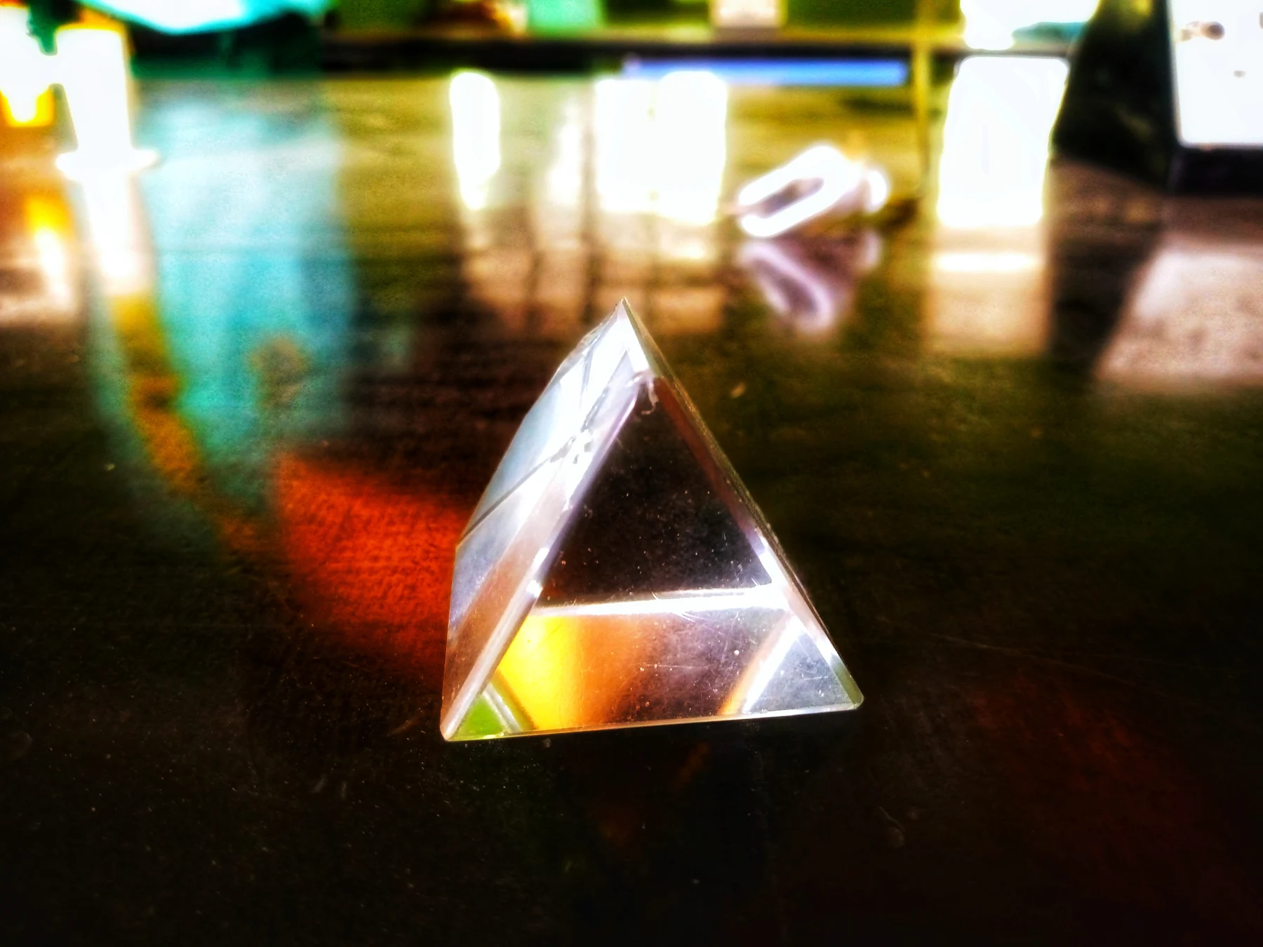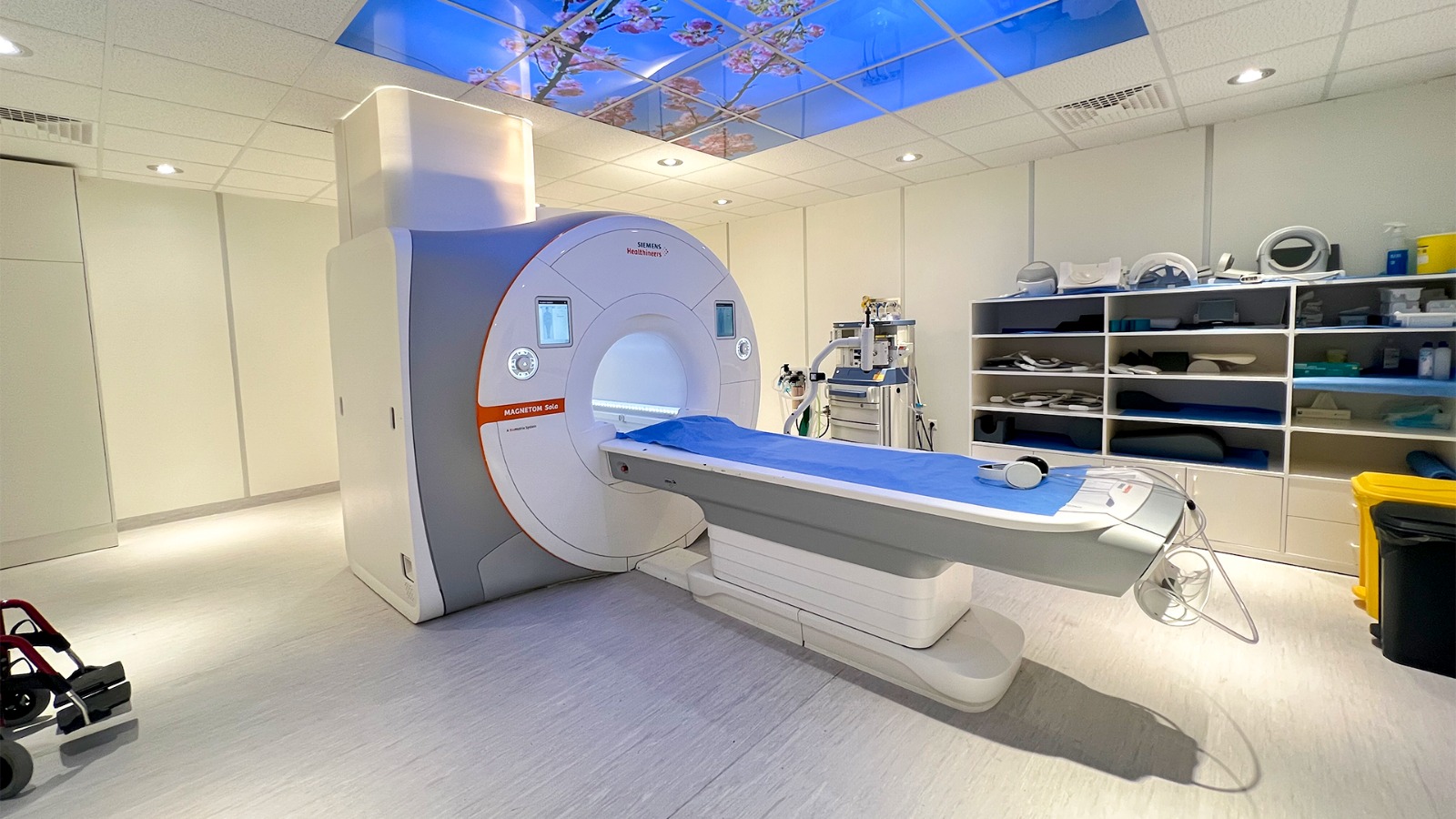
What is the difference between an electric field and a magnetic field, and why does medicine use both? An electric field is associated with voltage, while a magnetic field is associated with moving electric charge. Together they are part of electromagnetism, but they do not behave identically around materials or devices. MRI makes a useful example: it relies on a strong magnetic field and radiofrequency pulses to produce images, not on plasma or X-rays.
Voltage and current make different fields
An electric field describes how electric charge would experience a force in a region of space. Voltage is a difference in electrical potential, and greater voltage can produce a stronger electric field. A magnetic field is associated with moving charge, or current, and grows stronger as current increases. These are related ideas, but a static electric field can exist even when a device is off, while a device-generated magnetic field generally requires current to be flowing [1].
A simple contrast is a charged object and a current-carrying wire. The charged object can create an electric field around it; current in the wire creates a magnetic field. Magnetic fields can pass through many everyday materials, while electric fields are more readily weakened or shielded by walls and other objects [1]. The distinction matters in medicine because a field’s source, strength and interaction with the body depend on what is producing it and how the device operates.
Fields vary across the spectrum
Electromagnetic radiation spans a broad range of frequencies, from radio waves and visible light to X-rays and gamma rays. The higher-frequency end includes ionizing radiation, which has enough energy to damage DNA or cells directly; radiofrequency fields and visible light are in the non-ionizing portion [1]. “Radiation” therefore describes energy traveling or acting through space, not a single level of danger or a single biological effect.
Frequency is the number of oscillations per second. It is related to wavelength: higher-frequency waves have shorter wavelengths in a given medium. But frequency alone does not describe all exposure questions; intensity, distance, duration and the type of interaction also matter. MRI uses radiofrequency energy together with magnetic fields, and it does not use ionizing X-rays [2]. That is a precise distinction about the imaging method, not a claim that every electromagnetic field has the same effects or that any medical device is risk-free.
MRI turns proton behavior into contrast
In MRI, a powerful magnet aligns some of the protons in body water. A radiofrequency pulse then disturbs that alignment. When the pulse ends, protons move back toward alignment and release energy that the scanner detects. The timing and amount of that signal vary with the local molecular environment, allowing the system to distinguish tissues and build an image [2]. The scanner uses these magnetic responses to map anatomy rather than sending X-rays through the body.
A mental picture is a crowd of tiny spinning tops that have been nudged in a coordinated direction. The magnetic field provides the preferred direction; the radiofrequency pulse tips the tops away, and sensors detect their return. The analogy is simplified, but it captures why MRI needs both a strong magnetic field and carefully timed radiofrequency pulses. The signal depends on the environment around the protons, which is why different tissues can appear with different contrast [2].
The field is useful, but its setting matters
MRI’s lack of ionizing radiation does not mean the scanner contains no hazards. Its strong magnetic field can exert powerful forces on ferromagnetic objects, and patients need to disclose implants or devices so their compatibility can be assessed [2]. Scanners can also be loud, and rapidly switched fields may cause sensations such as twitching [2]. These are different issues from DNA damage by ionizing radiation; they arise from the physical properties and operation of the equipment.
More broadly, a field’s medical use should be understood by mechanism and evidence. MRI uses magnetic fields and radio waves; ultrasound uses sound; X-ray imaging uses ionizing electromagnetic radiation. Those technologies are not interchangeable simply because each can produce an image. NCI distinguishes non-ionizing fields from ionizing radiation and describes how electric and magnetic fields arise from voltage and current [1]. Knowing which field is present helps make sense of what a device measures, how it interacts with tissue and what safety questions are relevant.
Sources & further reading
- Electromagnetic Fields and Cancer National Cancer Institute
- Magnetic Resonance Imaging NIBIB
Prepared for the independent PSI Center reading library. About edition dates and source checking.
This explainer describes scientific principles. It does not assess individual treatment choices.