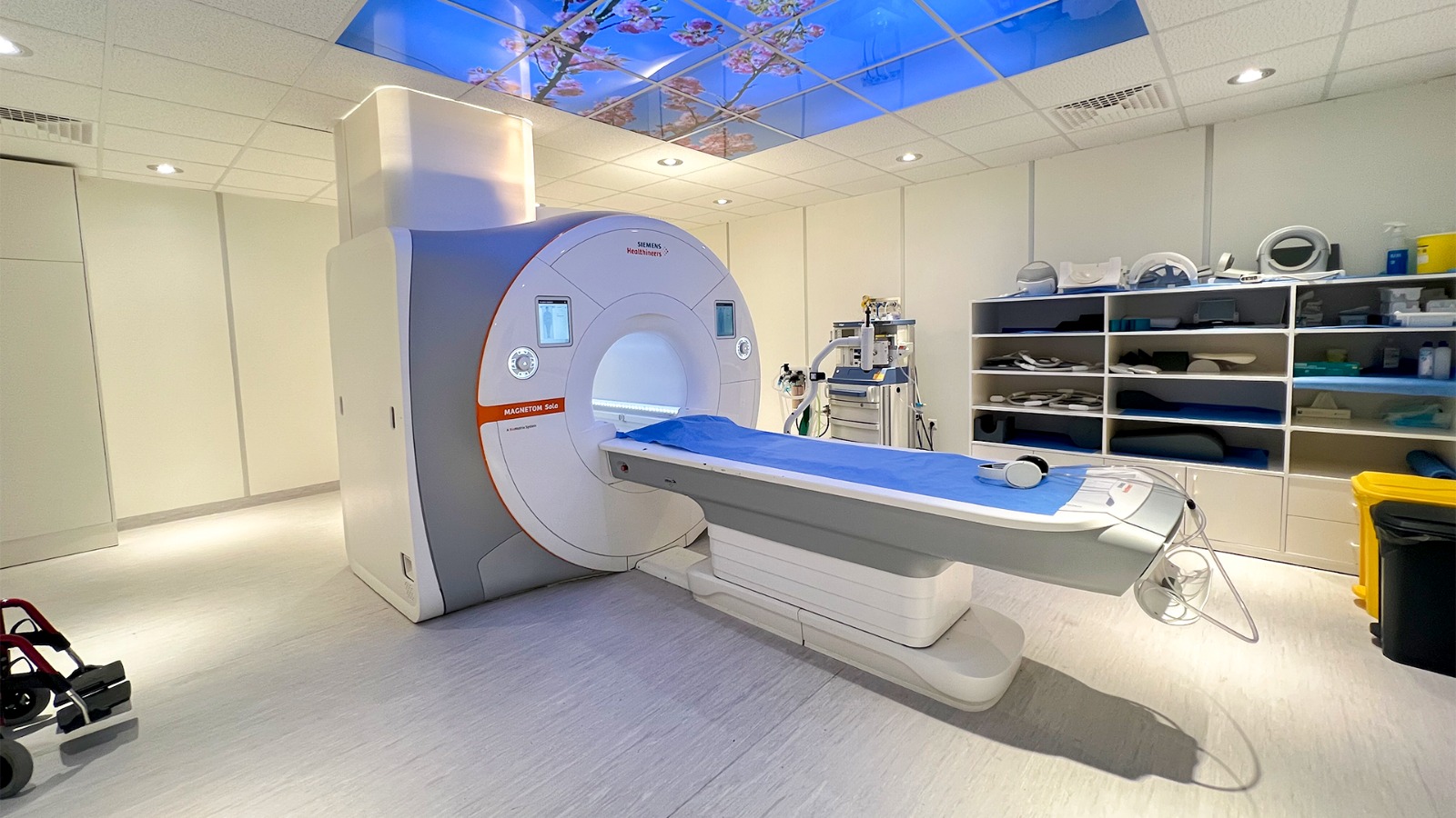
What links an MRI scanner’s radio pulse to the ear’s response to a musical note? Both involve systems responding to energy, but the mechanisms are quite different. In MRI, protons in body water are nudged by radiofrequency energy and their signals are measured. In hearing, sound moves fluid and tissue until sensory cells turn motion into electrical messages. Comparing them clarifies what resonance can—and cannot—mean.
A frequency is a repeating rhythm
A wave repeats a pattern in time or space. Its frequency tells how many cycles pass each second. Resonance occurs when a system responds especially strongly to energy delivered near one of its preferred frequencies. A swing offers a simple mental experiment: small pushes timed with its back-and-forth motion build amplitude more effectively than pushes delivered at random moments. The response depends on the system’s structure and the timing of the input.
The word “resonance” can describe different physical processes, so it should not blur their details. An MRI scanner does not listen to sound waves inside the body, and the ear does not use proton alignment. What connects them is a general pattern: a structured system responds to an input, and the resulting response contains clues about that system.
MRI reads proton behavior
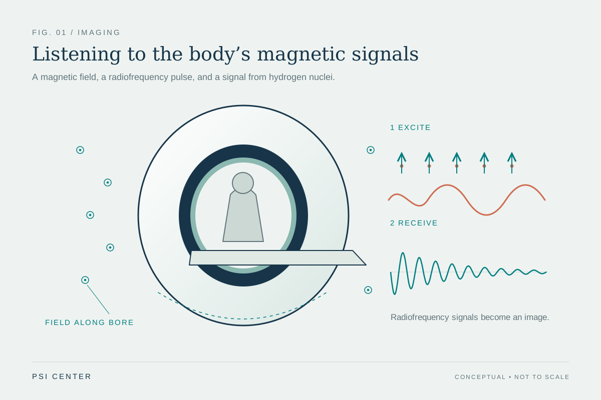
MRI uses a powerful magnetic field to align protons, especially those in water molecules, in the body. A radiofrequency pulse disturbs that alignment. When the pulse stops, the protons move back toward alignment and release energy that the scanner detects. The time course and amount of that signal vary with the molecules’ environment and chemical nature, allowing different tissues to produce different image contrast. [1]
The scanner combines these signals to create detailed anatomical images. It uses magnetic fields and radio waves, not ionizing x-rays. MRI is particularly useful for soft tissues such as the brain, spinal cord, muscles, ligaments, and tendons. The signal is not a direct photograph of a proton; it is measured energy processed into an image through the scanner’s physics and computation. [1]
The ear sorts motion by place
Hearing begins with sound waves traveling through the ear canal and vibrating the eardrum. Three small middle-ear bones transmit and amplify the vibration to the fluid-filled cochlea. There, a traveling wave moves along the basilar membrane. Hair cells ride this wave, and their position helps distinguish pitch: cells near the cochlea’s wide end respond to higher pitches, while those closer to its center respond to lower ones. [2]
As hair cells move, tiny projections called stereocilia bend against an overlying structure. That bending opens channels at their tips, allowing chemicals to enter and produce electrical signals. The auditory nerve carries those signals to the brain, which interprets them as sound. The ear’s frequency analysis emerges from anatomy and traveling motion, not from the magnetic alignment used in MRI. [2]
Two readouts, different kinds of information
Consider a steady musical tone and an MRI radiofrequency pulse as two inputs. The tone pushes air and then the eardrum, setting a chain of mechanical motion in motion; the cochlea’s position-dependent response helps encode pitch. The MRI pulse acts on proton spins in a magnetic field; the scanner measures the energy released as they realign. Both responses depend on the system’s surroundings, but one is a hearing pathway and the other an imaging signal.
This distinction helps explain why MRI can reveal anatomy without using x-rays, while hearing turns pressure changes in air into neural activity. MRI’s contrast depends on magnetic properties and proton relaxation; auditory pitch begins with sound-driven waves and hair-cell location. In each case, structure shapes the response, but the word “resonance” is not a license to treat the underlying processes as interchangeable. [1] [2]
Explore the physics: an explanatory diagram
Sources & further reading
- Magnetic Resonance Imaging NIBIB
- How Do We Hear? NIDCD
Prepared for the independent PSI Center reading library. About edition dates and source checking.
This explainer describes scientific principles. It does not assess individual treatment choices.