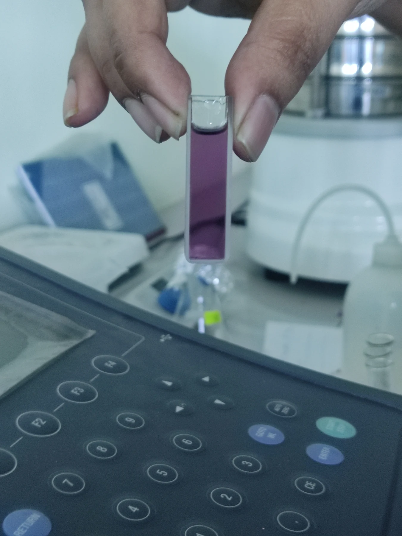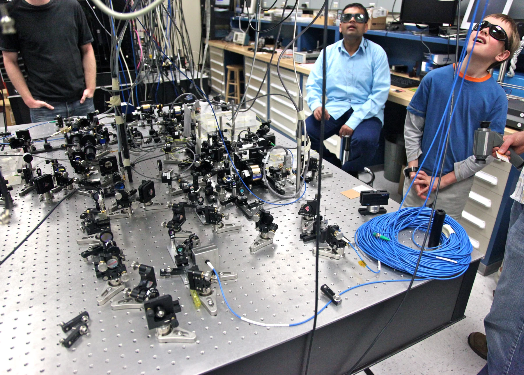
How can a glass slide help answer a question about disease inside a person? A microscope uses light and lenses to enlarge structures that are too small to see unaided. In medicine, a pathologist can examine cells and tissue architecture in a sample, looking for patterns that help guide diagnosis and treatment. The lens creates a view; careful interpretation connects that view to a clinical question.
Light becomes a magnified view
Optical imaging relies on the ways light interacts with objects: it can be absorbed, scattered, reflected, or emitted. Microscopy uses light and a system of lenses to magnify small objects, sometimes hundreds or thousands of times. A red blood cell, a cluster of cells, or details within a tissue sample becomes visible because the optical system gathers and focuses light into an image that the eye or camera can examine. [1]
Magnification alone is not the same as useful information. A larger image must still preserve enough detail and contrast for structures to be distinguished. Different parts of tissue interact with light differently, creating variations that make boundaries and patterns visible. This is why microscopy is more than “making small things big”: the instrument converts light from a specimen into a structured view that a trained observer can relate to biology.
From a slide to a clinical decision
Pathologists rely on microscopy images to diagnose disease and help guide treatment decisions. A tissue sample provides a limited window into the body: it represents the material collected, prepared, and placed under observation. The pathologist studies cell appearance and organization, then interprets those observations with the question being asked. Microscopy is foundational in medicine, but the image is evidence to be evaluated rather than an automatic diagnosis. [1]
Imagine two tissue samples that look similar at low magnification. Increasing magnification may reveal differences in cell arrangement or local structure that matter to interpretation. Conversely, a striking detail in one small field may not represent the whole specimen. This illustrates why diagnosis depends on both scale and sampling: the lens reveals fine features, while clinical reasoning asks how those features fit the broader pattern.
Devices extend what lenses can reach
Modern medical devices can monitor, modulate, or supplement body functions and structures. In optical medicine, cameras and fiber optics can carry imaging methods into internal cavities, allowing clinicians to visualize tissues in their natural setting. Other optical techniques use infrared light to image layers below a surface; optical coherence tomography, for example, is used to obtain detailed images within the retina. These approaches adapt the underlying interaction of light with tissue to different anatomical tasks. [2] [1]
A device’s role depends on its design and intended use. An endoscope combines a small camera with a route into an internal space; a microscope examines a specimen placed on a stage. Both produce visual information, but they answer different questions and face different constraints. An image from a device may support a clinical decision, yet it does not mean every optical tool diagnoses every condition or that all new imaging approaches are established practice.
More dimensions, more interpretation
A conventional tissue slide shows a thin section, so it offers a two-dimensional view of a sample that has depth and complexity. Researchers are developing methods to build and analyze three-dimensional pathology images. One challenge is that the resulting data can be too complex for manual review alone. A research platform described by NIBIB uses computational analysis to study 3D pathology images and predict outcomes, but such methods are not routinely analyzed in clinics today. [1]
The connection between a lens and diagnosis is therefore a chain: light interacts with tissue, optics form an image, a device captures or displays it, and a human or computational method interprets the pattern. Each link adds information and each has limits. Microscopy remains powerful because it makes otherwise inaccessible structure visible; diagnosis emerges when that view is weighed with the sample and the patient’s broader clinical picture. [2]
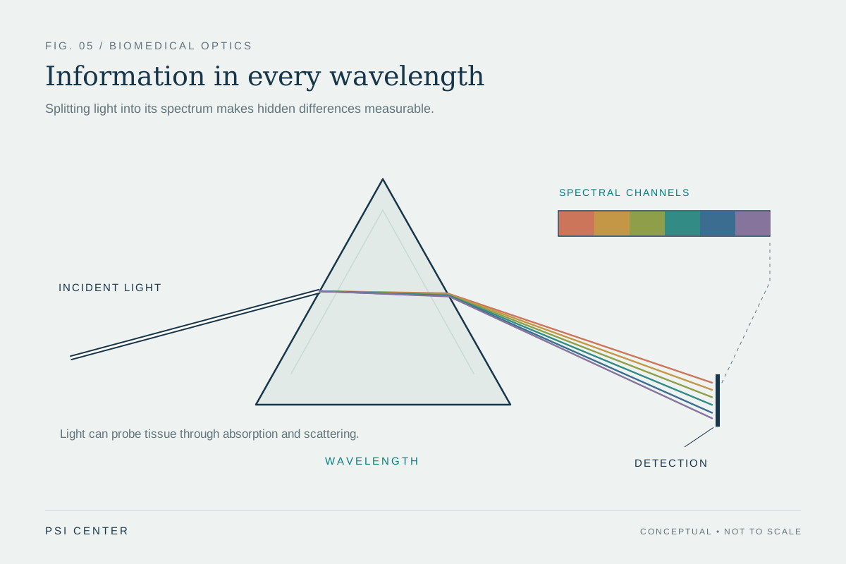
Explore the physics: an explanatory diagram
Sources & further reading
- Optical Imaging NIBIB
- Medical Device Technologies NIBIB
Prepared for the independent PSI Center reading library. About edition dates and source checking.
This explainer describes scientific principles. It does not assess individual treatment choices.