How can a clinician see the lining of a stomach without opening the abdomen? An endoscope carries light and a viewing system through a flexible instrument into a body cavity. Light illuminates tissue; some is reflected, absorbed, or scattered; and an optical path carries the returning signal to an eyepiece or camera. The resulting view is direct in an important sense, but it is still shaped by illumination, optics, motion, and the narrow route through which the instrument travels.
Light needs a route in and out
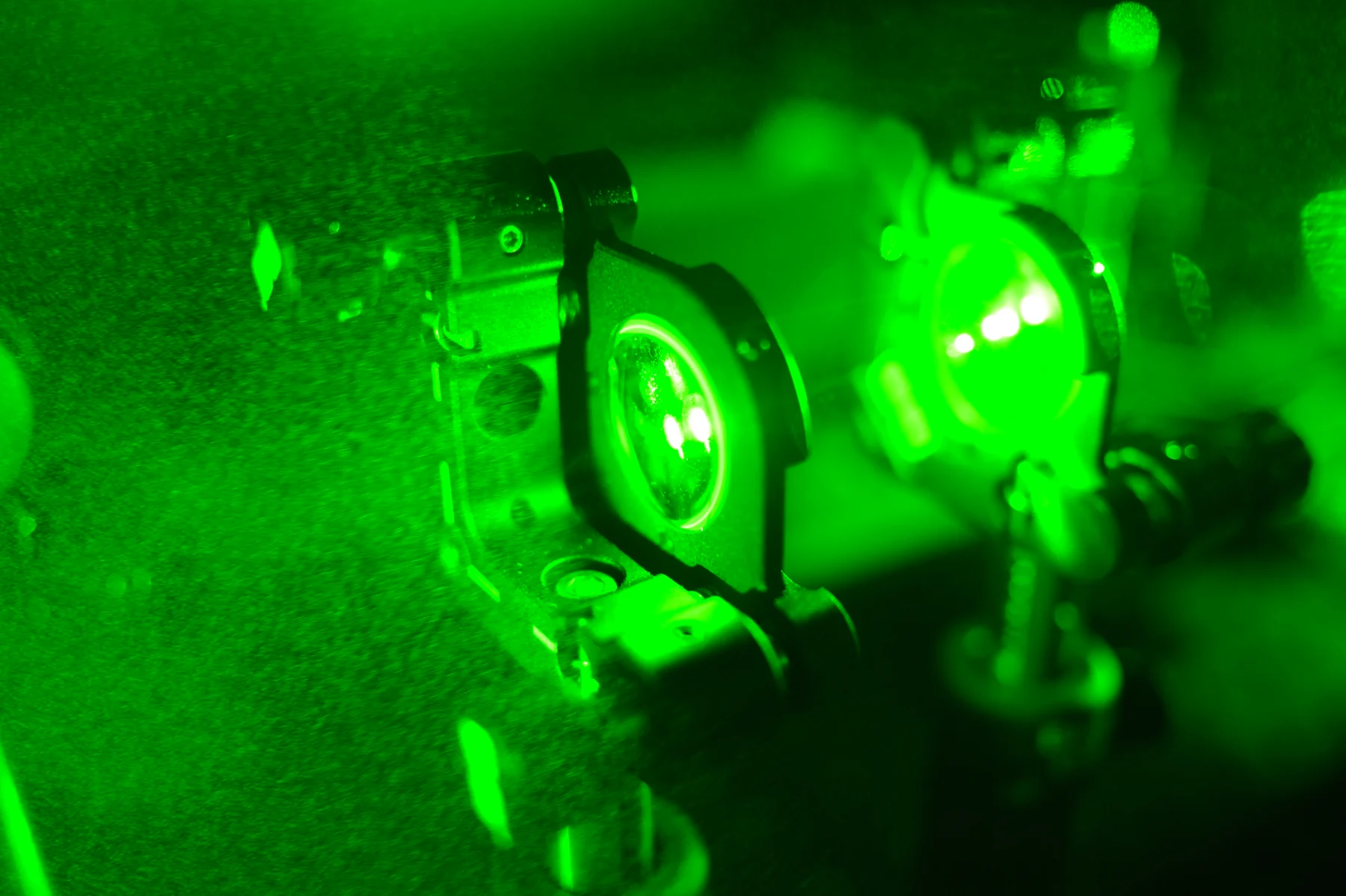
Optical imaging uses light’s interactions with matter to produce information about biological specimens. Tissue can absorb light, scatter it in many directions, reflect it, or emit light. In an endoscope, illumination must reach the tissue, and enough returning light must make its way back through the instrument to form a view. The narrow probe packages these functions so clinicians can inspect internal cavities such as the stomach. [1]
Fiber optics can carry light along flexible paths, while a miniature camera can capture an image near the end of an instrument. The exact arrangement varies among devices, but the design problem is similar: fit illumination and image collection into a slim tool that can bend or pass through anatomy. If the tissue is not lit, the camera receives little useful signal; if light is scattered or absorbed, contrast and visibility change.
What the picture represents
A familiar example is looking at a wet, curved surface with a small flashlight. The angle of the light changes which details stand out, while glare can obscure a patch. In a body cavity, tissue is more complex: its molecules and structures absorb and scatter wavelengths differently. The endoscopic view therefore reflects both anatomy and optical properties, and clinicians interpret the appearance rather than treating every color or brightness difference as a standalone diagnosis. [1]
Optical techniques can add detail beyond a conventional camera view. Optical coherence tomography uses infrared light to form cross-sectional images just below a surface, in a way that resembles ultrasound’s use of returning sound echoes. Other approaches combine light with ultrasound or measure near-infrared light to infer tissue properties. Endoscopy can adapt such optical methods to internal environments, but each technique answers different questions and has its own limits. [1]
A tool for seeing and acting
Endoscopes are not only viewing devices. Robotic systems can assist with endoscopic procedures, and specialized tools may allow clinicians to manipulate tissue while watching the procedure. The camera gives a view of the working area, while instruments provide access through a small route. This can support minimally invasive procedures, but the image is only one component: instrument control, anatomy, and clinical decisions also matter. [2]
The physical constraints are easy to overlook. A flexible tube must travel through a curved path, and its tip may not point in the direction the instrument body is moving. The operator works from the camera’s view, which can change as the scope turns or tissue moves. Robotic assistance can help with precision in some settings, and some systems provide high-definition three-dimensional views, but these are device capabilities rather than a promise that every endoscopic procedure is robotic or risk-free. [2]
The image has boundaries
Light generally does not travel through tissue as freely as it travels through a clear window. Absorption and scattering limit how deeply many optical methods can see, which helps explain why endoscopic viewing is useful in accessible cavities: the instrument brings the optics close to the surface being examined. Optical imaging can measure changes in soft tissue and may be combined with modalities such as MRI for additional information, but neither an optical image nor a camera view automatically settles a diagnosis. [1]
Medical devices include technologies that interact with the body to monitor, modulate, or support its functions. An endoscope fits this broad family, but devices differ greatly in purpose and evidence. Some optical methods are established tools, while others described in research settings are being developed. The useful question is what the particular instrument can show, how it obtains that signal, and how the image contributes to care. Seeing inside the body is an engineering achievement; interpreting what is seen remains a separate task. [2]
Sources & further reading
- Optical Imaging NIBIB
- Medical Device Technologies NIBIB
Prepared for the independent PSI Center reading library. About edition dates and source checking.
This explainer describes scientific principles. It does not assess individual treatment choices.