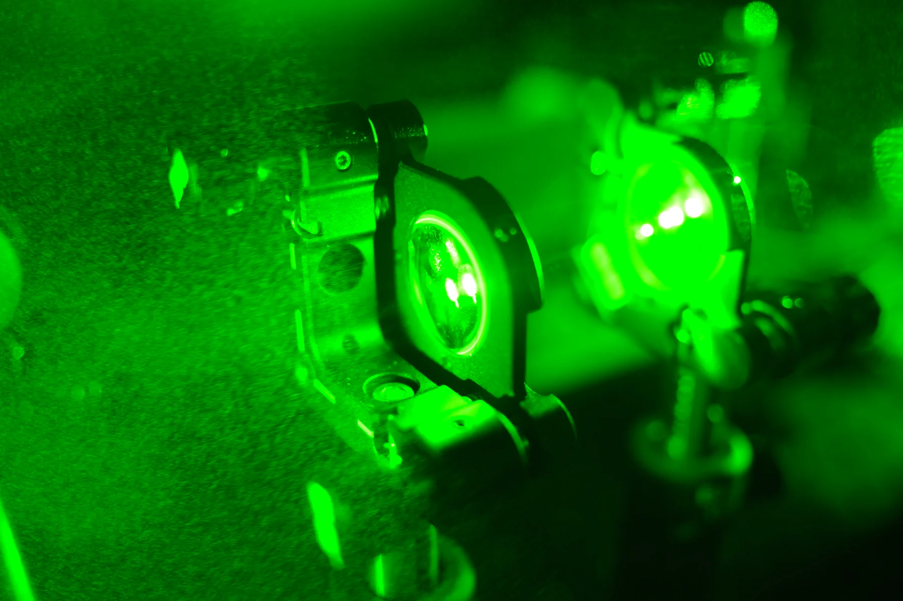
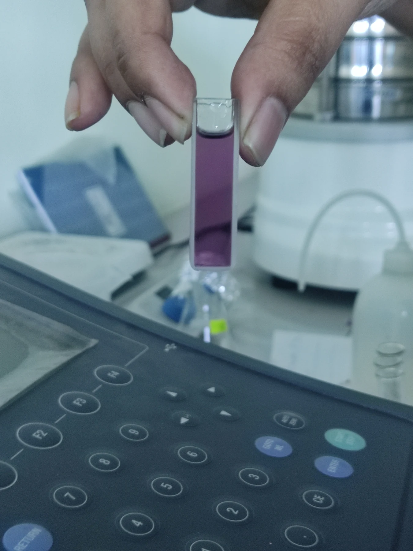
How can a microscope make one set of molecules stand out among thousands of neighboring structures? Fluorescence offers an answer: illuminate a sample with light that a chosen material absorbs, then detect the different light it emits. That color shift can make selected features easier to distinguish from their surroundings. The resulting image is not a neutral snapshot of everything present; it is a view shaped by what absorbs, emits, and can be detected.
Absorb, then emit
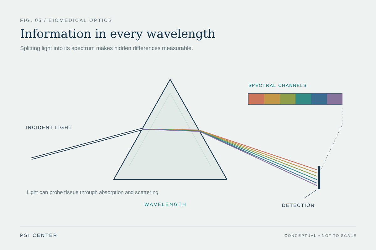
Light interacting with tissue can be absorbed, scattered, reflected, or emitted. In fluorescence, a molecule or label absorbs incoming light and then gives off light at a different wavelength, often a longer one. A microscope can illuminate a sample at the exciting wavelength and use filters to separate the emitted signal from reflected illumination. The contrast comes from the gap between those two bands of light.
A helpful mental experiment is to imagine a dark room with a patterned object painted in a fluorescent dye. Ordinary white light reveals the whole object; a chosen illumination and filter can make the dye’s pattern dominate the view. In biological samples, the “pattern” may correspond to a cell structure or molecule of interest. The detected light reveals where the signal is, not every detail of the surrounding biology. Optical imaging broadly uses light’s interactions with matter to visualize samples from molecules and cells to tissues and organisms [1].
Selection creates contrast
The method’s power is also its constraint. If a label attaches to a particular target, fluorescence can make that target easier to locate against a complex background. But the image only represents what the labeling strategy and optics can capture. A dark region might lack the target, the label, sufficient illumination, or a detectable signal. Brightness depends on more than how much biological material is present.
Microscopy is one part of optical imaging. Other techniques use light at larger scales or in different ways. Optical coherence tomography uses infrared light to create subsurface cross-sectional images, while diffuse optical methods use near-infrared light to measure properties such as hemoglobin concentration and oxygen saturation in soft tissue [1]. These methods do not all rely on fluorescence; their differences illustrate a broader point: the image depends on which interaction with light is being measured.
From cells to the body
Scale matters. Nanoscopic imaging can examine objects such as proteins or DNA strands; microscopic imaging resolves cells and smaller structures; macroscopic techniques can examine tissue samples or, in some cases, structures inside the body. A fluorescence image made from a thin laboratory sample is therefore not automatically comparable to a whole-organ view. At each scale, light must travel through material, and scattering can blur or weaken a signal.
Some optical imaging approaches are minimally or non-invasive, and light-based measurements can be repeated to follow changes over time. Optical methods can also be combined with other imaging techniques, including MRI, to add complementary information [1]. In a lab, fluorescence may answer where a selected signal appears. In a clinical setting, a method’s suitability depends on its particular design and purpose; the existence of a striking optical image by itself does not establish diagnostic value.
What the image leaves out
Fluorescence is best understood as a selective window. The label or naturally fluorescent molecule determines what can emit; the illumination determines what is excited; filters and detectors determine which photons count. Tissue absorption and scattering further influence what reaches the instrument. The image is thus a carefully constructed measurement, not a transparent view of the entire living system.
That distinction is useful when interpreting a vivid picture. A localized glow can support a claim about the presence or distribution of a detected signal under specified conditions. It cannot, on its own, prove that a disease is present, explain why a structure changed, or show that a treatment will help. Optical imaging supplies evidence by turning otherwise hard-to-see light interactions into spatial information; the biological meaning still depends on the question and the rest of the evidence [2].
Explore the physics: an explanatory diagram
Sources & further reading
- Optical Imaging NIBIB
- Rapid Diagnostics NIBIB
Prepared for the independent PSI Center reading library. About edition dates and source checking.
This explainer describes scientific principles. It does not assess individual treatment choices.