Why can visible light show the color of skin but not reveal the bones beneath it, while an x-ray image can? Both are electromagnetic radiation, waves of changing electric and magnetic fields. Their wavelengths and energies differ enough to produce very different interactions with matter. Light is absorbed, scattered, or reflected near the surface; x-rays can pass through the body, with tissues removing different fractions along the way.
One spectrum, different energies
Visible light and x-rays belong to the electromagnetic spectrum, but x-rays have much shorter wavelengths and higher photon energies. A photon is a packet of electromagnetic energy. When light reaches skin, molecules and structures in the tissue absorb and scatter parts of it, producing the colors and brightness our eyes detect. Optical imaging makes use of these interactions—absorption, scattering, reflection, or emitted light—to visualize specimens and tissues [1].
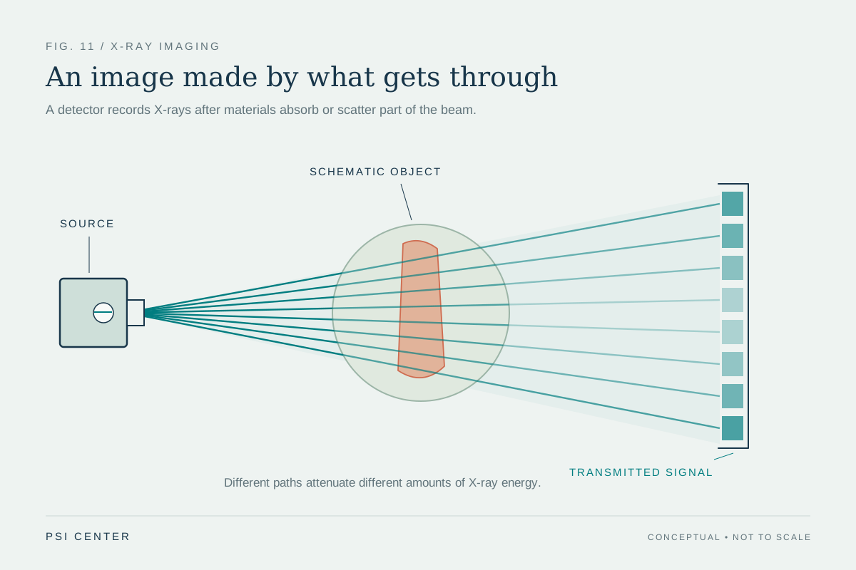
Higher-energy x-rays can penetrate tissue. Their transmission depends on how much is absorbed or scattered along a path. A detector records the remaining pattern, which can be processed into an image. Dense structures such as bone typically attenuate more x-rays than many soft tissues, creating contrast. Thus, the image is not a photograph of reflected light; it is a map of how much of an x-ray beam reached the detector [2].
Why the body creates contrast
Imagine shining a flashlight through a hand: visible light is scattered and absorbed so extensively that it does not produce a clear map of bones. In an x-ray image, however, the beam can traverse the hand, and differences in attenuation create shadows. Radiography records a single projection; computed tomography (CT) collects many x-ray views around the body and reconstructs cross-sectional slices [2].
Visible and infrared light can still reveal useful biological information. Optical coherence tomography uses infrared light to make cross-sectional images just below a surface, including detailed views of the retina. Photoacoustic imaging starts with laser light absorbed by tissue, which generates sound waves that a transducer detects. These methods show that light’s limited penetration can be a constraint for deep anatomy but an advantage when surface or tissue-property detail is the target [1].
What the image costs physically
X-rays are ionizing radiation: they have enough energy to potentially damage DNA. Medical x-ray imaging can provide valuable information for diagnosis, treatment planning, and procedures, but exposure carries a small potential increase in lifetime cancer risk that depends on factors such as dose, age, and body region [2]. That risk is one reason examinations should be justified by a medical question and optimized to use the lowest dose that still provides adequate image quality.
Visible, ultraviolet, and infrared light are less hazardous than x-rays in the contexts described for optical imaging, so optical techniques can be useful for repeated monitoring. But lower hazard does not mean every optical method can see deeply. Tissue absorbs and scatters light, limiting the distance it can travel in a clear, image-preserving way [1]. The signal that is easiest to use depends on whether the goal is surface detail, soft-tissue properties, or structures hidden deeper inside.
Choose the interaction that answers the question
The physics shapes the choice. A clinician looking for a broken bone may benefit from a radiograph because bone creates strong x-ray contrast. A specialist examining retinal layers can use optical coherence tomography because infrared light provides fine detail close to the eye’s surface. Neither method is a general-purpose window into the body; each measures a different interaction and produces evidence suited to particular questions [2] [1].
The distinction also helps avoid treating all radiation as equivalent. Visible light is non-ionizing and reaches the eye as a narrow range of electromagnetic wavelengths. X-rays are higher-energy and can ionize atoms, enabling penetration but also introducing radiation exposure considerations. A medical image is therefore a compromise between what the signal can reveal, how it travels through tissue, and what physical effects accompany its use.
Explore the physics: an explanatory diagram
Sources & further reading
- Optical Imaging NIBIB
- Medical X-ray Imaging FDA
Prepared for the independent PSI Center reading library. About edition dates and source checking.
This explainer describes scientific principles. It does not assess individual treatment choices.