What happens when a beam of light enters skin: does the tissue take it in, or send it off course? Both can happen, and they are not the same event. Absorption transfers light energy to matter; scattering redirects light through interactions with structures in the material. A detector sees the combined result. Separating these routes helps explain why optical imaging can reveal some tissue properties clearly while struggling to form a sharp picture deep beneath the surface.
Absorption transfers energy
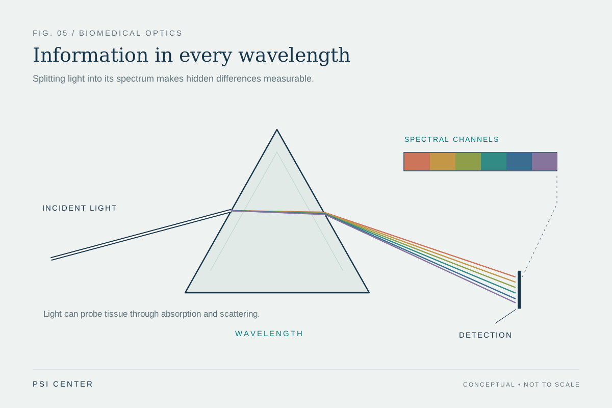
A photon is a packet of light energy. When a molecule absorbs a photon, the molecule takes up that energy, which may be converted into motion, heat, or a change in the molecule’s state. Different molecules absorb different wavelengths more readily. In tissue, this wavelength dependence can make light a probe of composition: blood pigments, for instance, affect how much light at particular wavelengths is absorbed [1].
Consider a flashlight shining through a colored filter. The filter appears colored because it absorbs some parts of the visible spectrum more than others, while other wavelengths pass through or return to the eye. Tissue is more complicated because it contains many absorbing molecules and layers, but the basic question remains: which wavelengths lose intensity, and by how much? Optical methods use such differences to estimate tissue properties [1].
Scattering redirects the path
Scattering changes a photon’s direction when it encounters variations in the material, such as cells, fibers, or boundaries. The light has not necessarily been absorbed; it may keep traveling, but along a new route. In tissue, many scattering events can turn an initially narrow beam into a diffuse glow. A detector positioned straight across from the source may then receive less light, even if the missing photons were redirected rather than absorbed [1].
This is why “the tissue is dark” is not a complete physical explanation for a dim signal. Absorption removes photons from the measured beam by transferring their energy; scattering can move photons away from a detector’s line of sight. Both reduce direct transmission, but they imply different interactions. Instruments can exploit or model the difference depending on whether they measure returned light, transmitted light, or light emerging from many directions [1].
A pulse can carry timing clues
Send a brief flash rather than a steady beam and the distinction gains another dimension. Light that travels a relatively direct route arrives earlier; photons that scatter repeatedly tend to take longer paths before reaching a detector. The timing and intensity of returning light can therefore contain information about how photons moved through tissue. This is a conceptual basis for using pulsed light to study biological materials, though real instruments must account for many overlapping paths [2].
Pulse oximeters offer a familiar example of light interacting with blood. A fingertip device sends red and near-infrared light through the finger and measures what reaches a detector on the other side. Oxygenated and deoxygenated blood absorb those wavelengths differently, so the device estimates oxygen saturation from their relative signals. The result depends on absorption, but also on a real measurement path through skin and tissue [2].
The tradeoff between depth and detail
Scattering helps explain why optical imaging often excels near surfaces and in thin or accessible tissues but faces challenges at depth. Repeated direction changes blur the relationship between where light entered and where it exits. Optical coherence tomography, for example, uses reflected infrared light to form sub-surface images, including detailed views within the retina. Photoacoustic imaging takes another route: absorbed laser pulses generate sound waves that can be detected to map deeper tissue [1].
No single detector reading cleanly reports one process in isolation. A change in signal may reflect a change in absorber concentration, scattering structure, geometry, or several at once. Researchers choose wavelengths, pulse timing, and detector arrangements to constrain that interpretation. Absorption and scattering are distinct physical routes, but in living tissue they work together to set what a light-based instrument can see [1] [2].
Explore the physics: an explanatory diagram
Sources & further reading
- Optical Imaging NIBIB
- Updating Pulse Oximeters NHLBI
Prepared for the independent PSI Center reading library. About edition dates and source checking.
This explainer describes scientific principles. It does not assess individual treatment choices.